Miễn phí
Viêm cơ hoại tử do trung gian MDA5 liên quan đến COVID-19
COVID-19-Associated MDA5-Mediated Necrotizing Myositis
Sarah Steadman; Amit Sikder; Harsh R. Desai; Bradley Collins; Tiffany Baker; Julie Worthington. American Journal of Case Reports. doi: 10.12659/AJCR.949788. Open access.
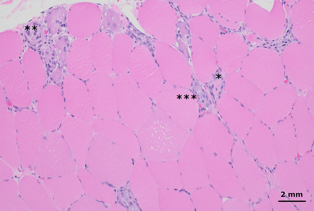
Figure 1
https://jours.isi-science.com/imageXml.php?i=amjcaserep-26-e949788-g001.jpg&idArt=949788&w=1000
Figure 1
Hình 1
Hematoxylin and eosin-stained sections from formalin-fixed paraffin-embedded skeletal muscle tissue shows myofibers at different stages of degeneration and regeneration, including necrosis (*), myophagocytosis (**), and basophilic fibers (*** left of basophilic fibers). These changes affected a minority of the muscle tissue and were diffusely distributed throughout the muscle fascicles, without an obvious perifascicular pattern. There was no significant lymphocytic inflammation (200× total magnification).
Các lát cắt nhuộm hematoxylin và eosin từ mô cơ vân cố định bằng formalin và đúc trong paraffin cho thấy các sợi cơ ở những giai đoạn thoái hóa và tái tạo khác nhau, bao gồm hoại tử (*), hiện tượng thực bào cơ (**), và các sợi cơ bắt màu kiềm (*** phía bên trái của các sợi cơ bắt màu kiềm). Những thay đổi này gây ảnh hưởng đến một phần nhỏ mô cơ và phân bố lan tỏa khắp các bó cơ, không có dạng tổn thương quanh bó cơ rõ rệt. Không ghi nhận tình trạng viêm nhiễm lymphocytic đáng kể (độ phóng đại tổng cộng 200×).
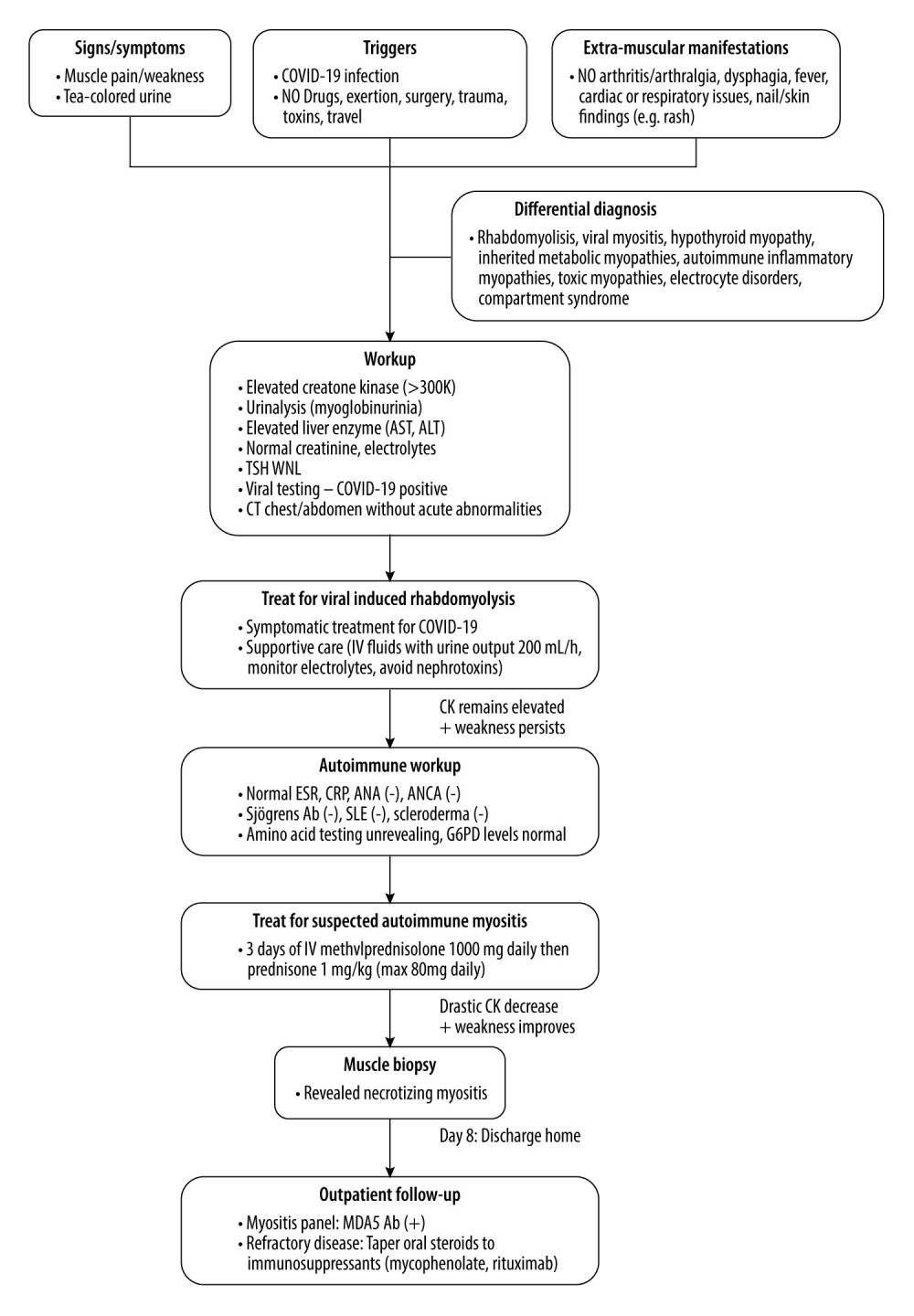
Figure 2
https://jours.isi-science.com/imageXml.php?i=amjcaserep-26-e949788-g002.jpg&idArt=949788&w=1000
Figure 2
Hình 2
Flow diagram illustrating the decision-making process in diagnosing and managing this case.
Sơ đồ dòng mô tả quy trình ra quyết định trong chẩn đoán và xử trí ca bệnh này.
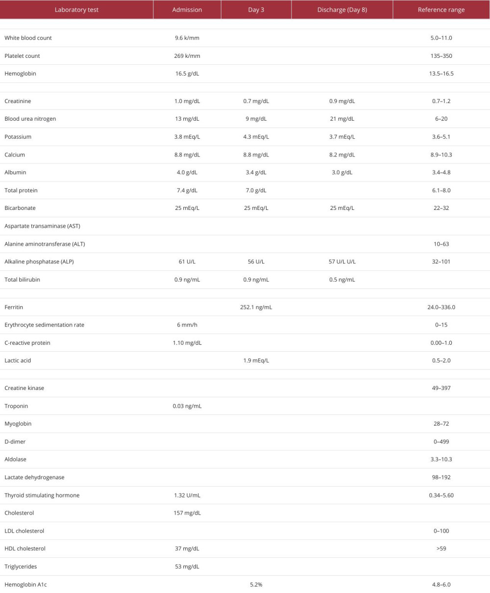
Table 1
https://jours.isi-science.com/imageXml.php?i=t1-amjcaserep-26-e949788.jpg&idArt=949788&w=1000
Table 1
Bảng 1
Biochemical laboratory test values during hospital admission.
Giá trị các xét nghiệm hóa sinh trong thời gian nằm viện.
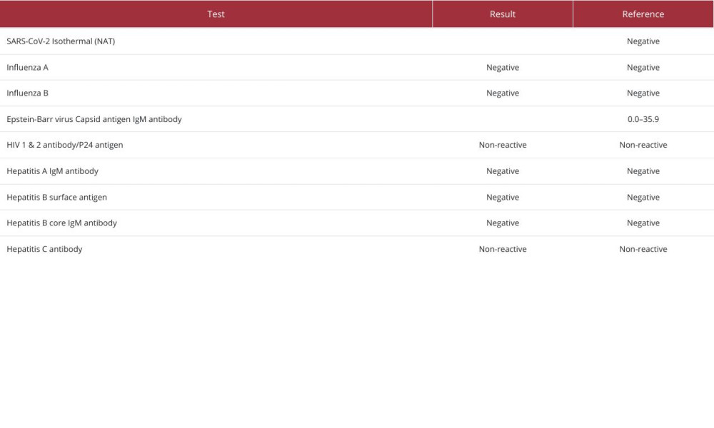
Table 2
https://jours.isi-science.com/imageXml.php?i=t2-amjcaserep-26-e949788.jpg&idArt=949788&w=1000
Table 2
Bảng 2
Test results for viral infections during hospital admission.
Kết quả xét nghiệm các tình trạng nhiễm virus trong thời gian nằm viện.
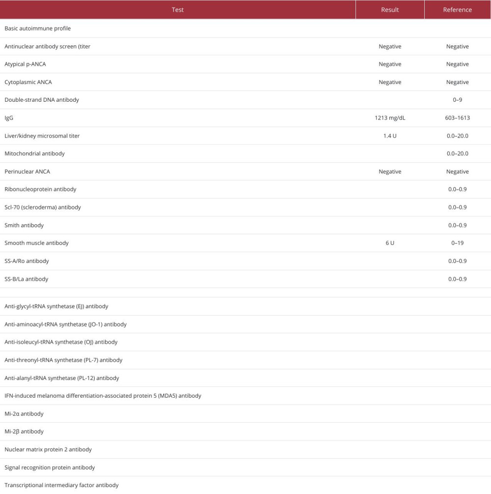
Table 3
https://jours.isi-science.com/imageXml.php?i=t3-amjcaserep-26-e949788.jpg&idArt=949788&w=1000
Table 3
Bảng 3
Autoimmune testing performed during hospital admission and outpatient follow-up.
Các xét nghiệm tự miễn được thực hiện trong thời gian nằm viện và theo dõi ngoại trú.
Có thể bạn quan tâm
-
Mang thai sau khi hóa trị điều trị ung thư biểu mô màng đệm trong nhau thai: Theo dõi 5 năm thuận lợi đối với mẹ và con
Conception After Chemotherapy for Intraplacental Choriocarcinoma: Favorable 5-Year Follow-Up of the Mother and Child
-
Tỷ lệ sống còn toàn bộ với relacorilant và nab-paclitaxel ở bệnh nhân ung thư buồng trứng kháng platinum (ROSELLA): một thử nghiệm kiểm chứng ngẫu nhiên pha 3
Overall survival with relacorilant and nab-paclitaxel in patients with platinum-resistant ovarian cancer (ROSELLA): a phase 3 randomised controlled trial
-
Virus Marburg lây lan giữa các loài như thế nào? Ảnh của nhà khoa học trẻ người Uganda cung cấp manh mối quan trọng
How does Marburg virus spread between species? Young Ugandan scientist’s photos give important clues