Miễn phí
U tủy xương ác tính nguyên phát ở phổi biểu hiện dưới dạng nốt dưới màng phổi kích thước 1.7 cm ở nam bệnh nhân 61 tuổi: báo cáo ca lâm sàng
Primary Pulmonary Osteosarcoma Presenting as a 1.7-cm Subpleural Nodule in a 61-Year-Old Man: A Case Report
Xin Yang; Wen Guang Zhao; Chao Gao; Sheng Lin. American Journal of Case Reports. doi: 10.12659/AJCR.949034. Open access.
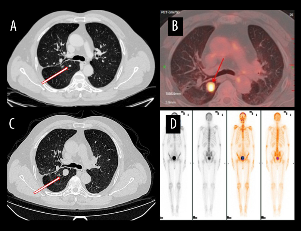
Figure 1
https://jours.isi-science.com/imageXml.php?i=amjcaserep-26-e949034-g001.jpg&idArt=949034&w=1000
Figure 1
Hình 1
Chest imaging workup of the right upper lobe nodule(A) Computed tomography (CT) from August 2024 shows a 0.7-cm, smooth-edged subpleural nodule. (B) CT from January 2025 demonstrates interval growth to 1.7 cm. (C) Positron-emission tomography/CT reveals increased uptake (SUVmax 5.2). (D) Whole-body bone scintigraphy shows no skeletal uptake, providing evidence against a bone primary or osseous metastasis.
Khảo sát hình ảnh nốt thùy trên phổi phải (A) Chụp cắt lớp vi tính (CT) tháng 8 năm 2024 cho thấy một nốt dưới màng phổi bờ nhẵn kích thước 0,7 cm. (B) CT tháng 1 năm 2025 cho thấy tổn thương tăng kích thước lên 1,7 cm theo thời gian. (C) Chụp cắt lớp phát xạ positron/CT cho thấy tăng hấp thu (SUVmax 5,2). (D) Xạ hình xương toàn thân không ghi nhận tăng hấp thu tại hệ xương, cung cấp bằng chứng chống lại nguồn gốc nguyên phát từ xương hoặc di căn xương.
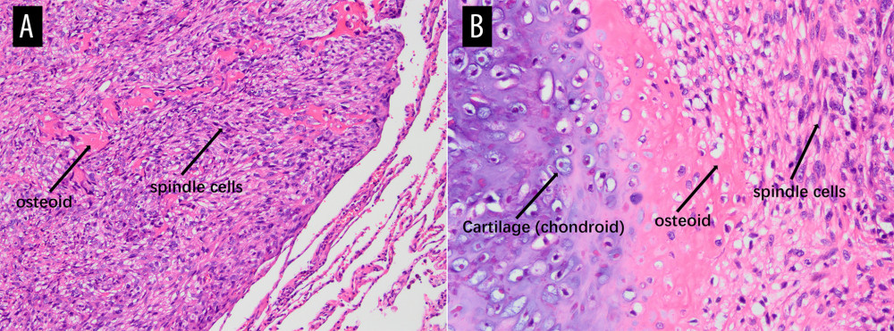
Figure 2
https://jours.isi-science.com/imageXml.php?i=amjcaserep-26-e949034-g002.jpg&idArt=949034&w=1000
Figure 2
Hình 2
Histopathology of primary pulmonary osteosarcoma with hematoxylin and eosin staining(A) Magnification ×100. Low-power view of a hypercellular spindle-cell neoplasm within lung parenchyma, with irregular dense eosinophilic osteoid deposits (arrow named “osteoid”) rimmed and permeated by tumor cells (labeled “spindle cells”). Residual alveolar septa/fibrous stroma are present at the periphery, indicating adjacency to native lung. (B) Magnification ×200. Higher-power view demonstrates a triphasic pattern with focal chondroid differentiation (labeled “Cartilage, chondroid”) showing basophilic matrix containing lacunae with chondrocyte-like tumor cells, transitioning to abundant lace-like/trabecular eosinophilic osteoid (labeled “osteoid”). The osteoid is directly produced by and rimmed and permeated by atypical spindle tumor cells (labeled “spindle cells”), exhibiting nuclear pleomorphism, hyperchromasia, and frequent mitotic figures. The intimate association of malignant spindle cells with tumor osteoid, together with focal chondroid differentiation, supports an osteoid-forming high-grade sarcoma (osteoblastic osteosarcoma phenotype).
Mô bệnh học của osteosarcoma phổi nguyên phát nhuộm hematoxylin và eosin (A) Độ phóng đại ×100. Hình ảnh công suất thấp cho thấy một khối u tế bào hình thoi tăng mật độ nằm trong nhu mô phổi, với các lắng đọng osteoid ái eosin đậm đặc không đều (mũi tên ký hiệu “osteoid”) được viền quanh và thấm nhập bởi các tế bào u (ký hiệu “spindle cells”). Các vách phế nang/mô đệm xơ còn sót lại hiện diện ở ngoại vi, cho thấy tổn thương nằm kế cận mô phổi bản địa. (B) Độ phóng đại ×200. Hình ảnh công suất cao hơn cho thấy kiểu hình ba pha với biệt hóa dạng sụn khu trú (ký hiệu “Cartilage, chondroid”) biểu hiện chất nền ái kiềm chứa các ổ khuyết với các tế bào u giống tế bào sụn, chuyển tiếp sang các dải osteoid ái eosin dạng ren/dạng bè phong phú (ký hiệu “osteoid”). Osteoid được tạo trực tiếp, được viền quanh và thấm nhập bởi các tế bào u hình thoi không điển hình (ký hiệu “spindle cells”), biểu hiện đa dạng nhân, tăng sắc và nhiều hình ảnh phân bào. Mối liên quan mật thiết giữa các tế bào hình thoi ác tính với osteoid của khối u, cùng với biệt hóa dạng sụn khu trú, hỗ trợ chẩn đoán sarcoma độ cao tạo osteoid (kiểu hình osteosarcoma tạo xương).
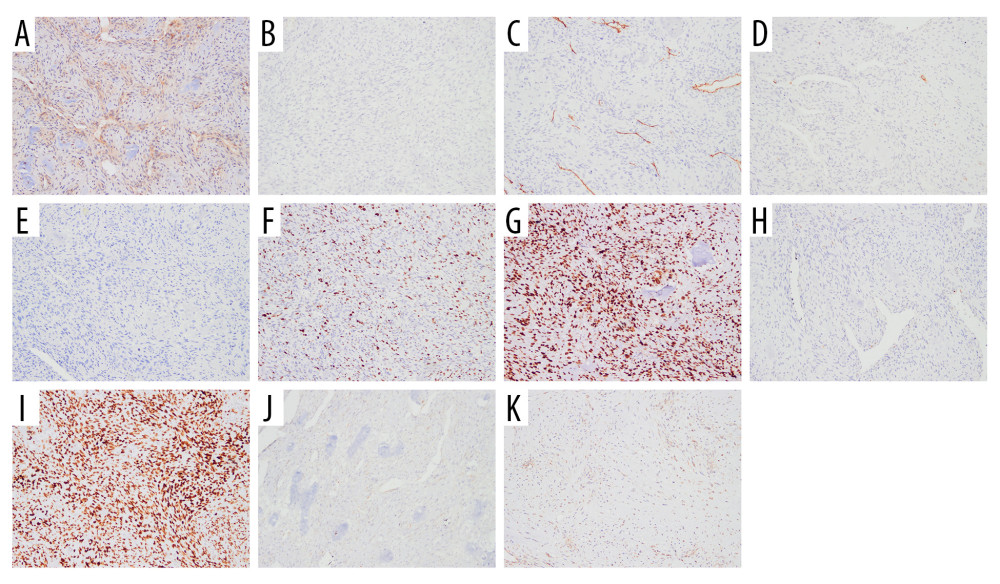
Figure 3
https://jours.isi-science.com/imageXml.php?i=amjcaserep-26-e949034-g003.jpg&idArt=949034&w=1000
Figure 3
Hình 3
Immunohistochemical panel of the tumor (all with magnification ×100)(A) B-cell lymphoma 2 (BCL-2): focal positivity. (B) Low-molecular-weight cytokeratin antibody CK8/18 (CAM5.2): negative (rare weak-positive cells). (C) CD34: restricted to vascular endothelium (internal control). (D) Cytokeratin: negative (rare weak-positive cells). (E) Desmin: negative. (F) Ki-67 proliferation: approximately 30% labeling index. (G) Tumor suppressor protein (p53): diffuse strong nuclear staining (mutant-type pattern). (H) S100 protein family: focal positivity. (I) Special AT-rich sequence-binding protein 2 (SATB2): diffuse nuclear positivity. (J) Smooth muscle actin (SMA): focal positivity. (K) Signal transducer and activator of transcription 6 (STAT6): negative. Epithelial membrane antigen (not shown) was also negative. The overall IHC profile supports osteoblastic differentiation and argues against epithelial, vascular, and solitary fibrous tumor lineages.
Bảng nhuộm hóa mô miễn dịch của khối u (tất cả ở độ phóng đại ×100) (A) B-cell lymphoma 2 (BCL-2): dương tính khu trú. (B) Kháng thể cytokeratin trọng lượng phân tử thấp CK8/18 (CAM5.2): âm tính (hiếm tế bào dương tính yếu). (C) CD34: chỉ giới hạn ở nội mô mạch máu (đối chứng nội tại). (D) Cytokeratin: âm tính (hiếm tế bào dương tính yếu). (E) Desmin: âm tính. (F) Chỉ số tăng sinh Ki-67: khoảng 30%. (G) Protein ức chế khối u (p53): nhuộm nhân mạnh lan tỏa (kiểu đột biến). (H) Họ protein S100: dương tính khu trú. (I) Special AT-rich sequence-binding protein 2 (SATB2): dương tính nhân lan tỏa. (J) Smooth muscle actin (SMA): dương tính khu trú. (K) Signal transducer and activator of transcription 6 (STAT6): âm tính. Kháng nguyên màng biểu mô (không hiển thị) cũng âm tính. Hồ sơ hóa mô miễn dịch tổng thể hỗ trợ biệt hóa tạo xương và chống lại nguồn gốc biểu mô, mạch máu và u xơ đơn độc.
Có thể bạn quan tâm
-
U bướu thần kinh nội tiết biệt hóa tốt nguyên phát ở vú: Báo cáo ca bệnh và tổng quan tài liệu
Primary Well-Differentiated Neuroendocrine Tumor of the Breast: A Case Report and Literature Review
-
Miếng dán Estradiol qua da trong điều trị ung thư tuyến tiền liệt tiến triển tại chỗ
Transdermal Estradiol Patches in Locally Advanced Prostate Cancer
-
Dị ứng phấn hoa năm nay rất nặng – bác sĩ giải thích lý do, và cách tìm cách giảm nhẹ
Pollen allergies are brutal this year – a doctor explains why, and how to find relief