Ca lâm sàng
U bướu thần kinh nội tiết biệt hóa tốt nguyên phát ở vú: Báo cáo ca bệnh và tổng quan tài liệu
Primary Well-Differentiated Neuroendocrine Tumor of the Breast: A Case Report and Literature Review
Sanjna Sai Gudena; Nilesh Digvijay Kashikar; Matthew Charles Wakefield. American Journal of Case Reports. doi: 10.12659/AJCR.950684. Open access.
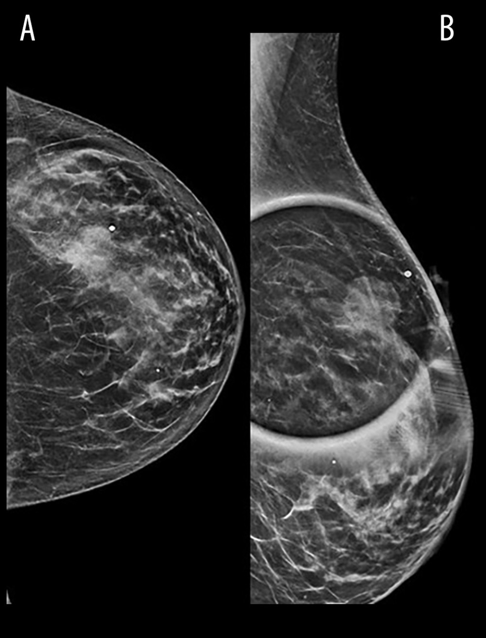
Figure 1
https://jours.isi-science.com/imageXml.php?i=amjcaserep-26-e950684-g001.jpg&idArt=950684&w=1000
Figure 1
Hình 1
(A) Mammogram revealing a 2.6-cm mass in the left breast upper outer quadrant. (B) CC view confirming the breast mass.
(A) Phim chụp X-quang tuyến vú (mammogram) cho thấy một khối u kích thước 2,6 cm ở phần tư trên ngoài của vú trái. (B) Hình ảnh tư thế CC (craniocaudal) xác nhận khối u ở vú.
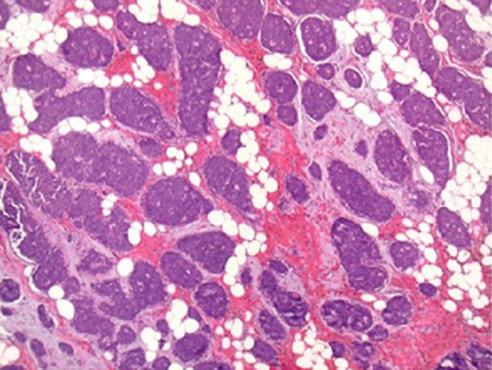
Figure 2
https://jours.isi-science.com/imageXml.php?i=amjcaserep-26-e950684-g002.jpg&idArt=950684&w=1000
Figure 2
Hình 2
Low-magnification view (40×) of the tumor with H&E stain, showing tumor cells arranged in tight organoid nests with delicate intervening fibrovascular stroma. Almost the entire tumor shows the same pattern.
Hình ảnh độ phóng đại thấp (40×) của khối u được nhuộm H&E, cho thấy các tế bào u sắp xếp thành các ổ dạng cơ quan (organoid) chặt chẽ với mô đệm xơ mạch mỏng xen kẽ. Hầu như toàn bộ khối u đều thể hiện mô hình tương tự.
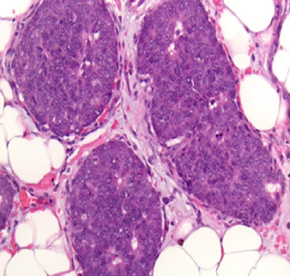
Figure 3
https://jours.isi-science.com/imageXml.php?i=amjcaserep-26-e950684-g003.jpg&idArt=950684&w=1000
Figure 3
Hình 3
At a higher magnification (200×), the tumor cells show many typical features of neuroendocrine morphology. The tumor cells are polygonal with granular eosinophilic cytoplasm. The classical salt-and-pepper stippled chromatin is visible in a minority of tumor cells. Neuroendocrine tumors of the breast in general, as also seen in this case, tend to show a lower Nottingham nuclear grade of 1 or 2 out of 3.
Ở độ phóng đại cao hơn (200×), các tế bào u thể hiện nhiều đặc điểm hình thái điển hình của thần kinh nội tiết. Các tế bào u có hình đa giác với tế bào chất dạng hạt ưa eosin. Chất nhiễm sắc lốm đốm kiểu “muối tiêu” cổ điển có thể nhìn thấy ở một số ít tế bào u. Nhìn chung, các u thần kinh nội tiết ở vú, và cũng được ghi nhận trong trường hợp này, có xu hướng thể hiện độ nhân Nottingham thấp hơn, ở mức 1 hoặc 2 trên 3.
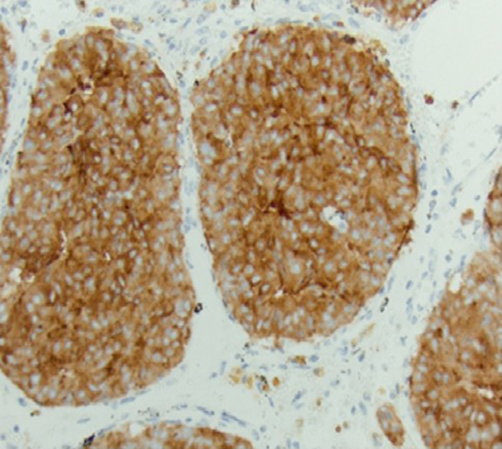
Figure 4
https://jours.isi-science.com/imageXml.php?i=amjcaserep-26-e950684-g004.jpg&idArt=950684&w=1000
Figure 4
Hình 4
Immunohistochemical stain for synaptophysin, a typical marker for neuroendocrine differentiation, displays cytoplasmic labeling within the tumor cells, confirming the neuroendocrine nature of the tumor.
Nhuộm hóa mô miễn dịch đối với synaptophysin, một dấu ấn điển hình cho sự biệt hóa thần kinh nội tiết, cho thấy sự gắn nhãn tế bào chất bên trong các tế bào u, xác nhận bản chất thần kinh nội tiết của khối u.
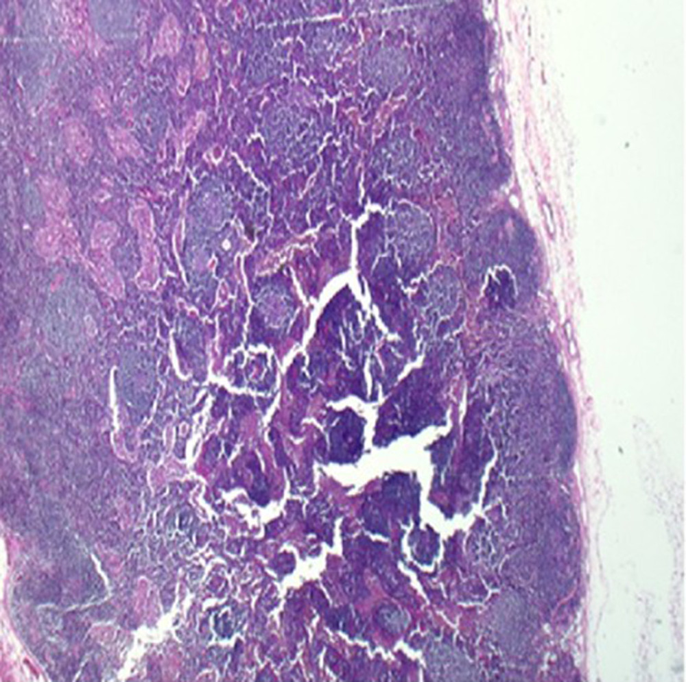
Figure 5
https://jours.isi-science.com/imageXml.php?i=amjcaserep-26-e950684-g005.jpg&idArt=950684&w=1000
Figure 5
Hình 5
A representative micrograph from one of the lymph nodes showing normal lymph node architecture with lymphoid follicles and sinusoids, without any evidence of metastatic tumor.
Hình ảnh vi thể đại diện từ một trong các hạch bạch huyết cho thấy cấu trúc hạch bạch huyết bình thường với các nang bạch huyết và xoang hạch, không có bất kỳ bằng chứng nào của u di căn.
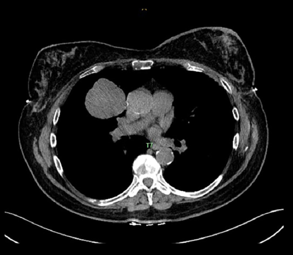
Figure 6
https://jours.isi-science.com/imageXml.php?i=amjcaserep-26-e950684-g006.jpg&idArt=950684&w=1000
Figure 6
Hình 6
Computed tomography showing the right upper-lobe lung mass and mediastinal lymphadenopathy.
Chụp cắt lớp vi tính cho thấy khối u ở thùy trên phổi phải và bệnh lý hạch bạch huyết trung thất.
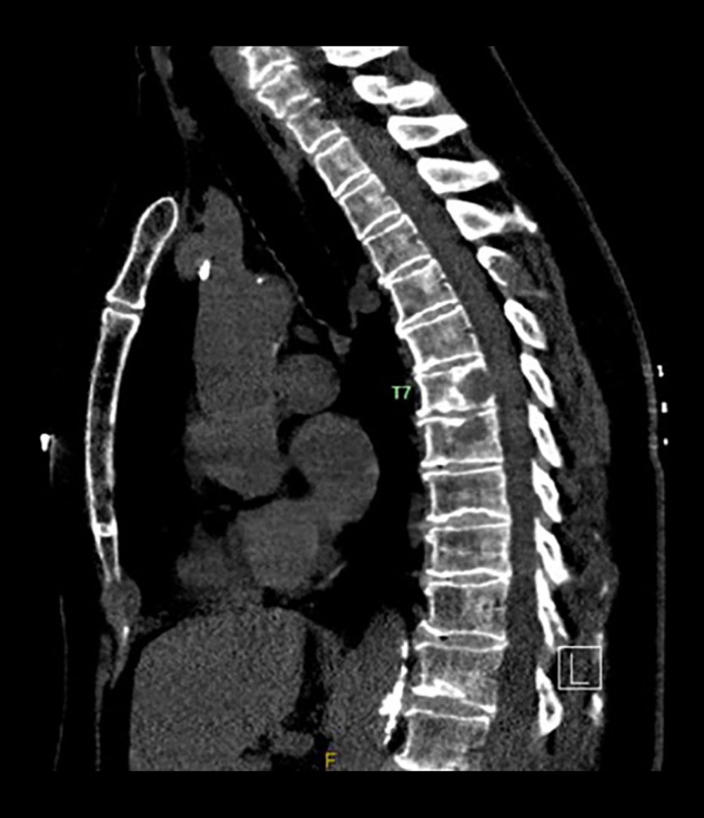
Figure 7
https://jours.isi-science.com/imageXml.php?i=amjcaserep-26-e950684-g007.jpg&idArt=950684&w=1000
Figure 7
Hình 7
Computed tomography of the thoracic spine showing a T7 vertebral compression fracture.
Chụp cắt lớp vi tính cột sống ngực cho thấy xẹp đốt sống T7 do gãy xương bệnh lý.
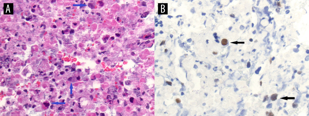
Figure 8
https://jours.isi-science.com/imageXml.php?i=amjcaserep-26-e950684-g008.jpg&idArt=950684&w=1000
Figure 8
Hình 8
Lung involvement by metastatic breast carcinoma. (A) High-power magnification image of the H&E-stained fine-needle aspirate from the lung lesion appears to be predominantly necrotic with an admixture of inflammatory cells, macrophages, and a few probable metastatic malignant cells (blue arrows). (B) Immunohistochemical stain for a breast marker, GATA3, was performed. The stain labeled rare, scattered, malignant-appearing cells (black arrows), confirming the presence of metastatic breast carcinoma to the lung.
Sự xâm lấn phổi do ung thư biểu mô tuyến vú di căn. (A) Hình ảnh độ phóng đại cao của bệnh phẩm chọc hút bằng kim nhỏ từ tổn thương phổi được nhuộm H&E cho thấy vùng hoại tử là chủ yếu xen lẫn các tế bào viêm, đại thực bào và một vài tế bào ác tính có khả năng là di căn (mũi tên màu xanh). (B) Nhuộm hóa mô miễn dịch đối với một dấu ấn của vú, GATA3, đã được thực hiện. Thuốc nhuộm đã gắn nhãn các tế bào hiếm gặp, rải rác, có biểu hiện ác tính (mũi tên màu đen), xác nhận sự hiện diện của ung thư biểu mô tuyến vú di căn đến phổi.
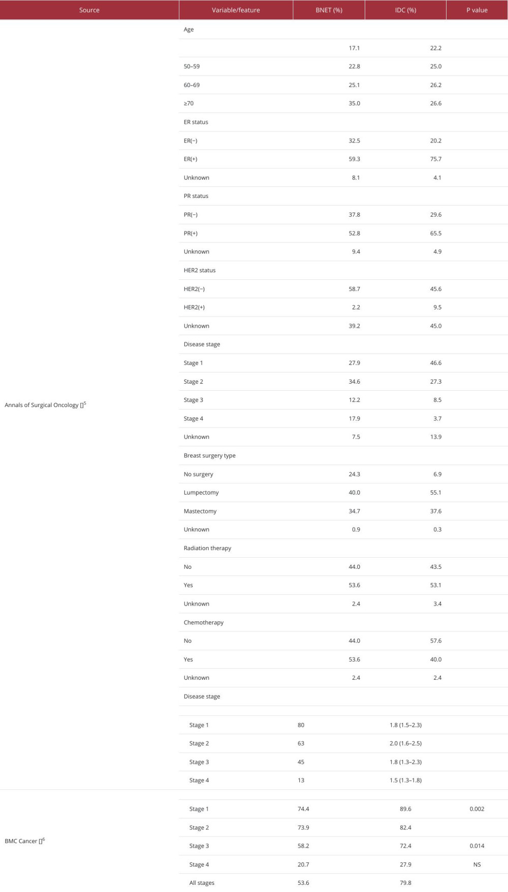
Table 1
https://jours.isi-science.com/imageXml.php?i=t1-amjcaserep-26-e950684.jpg&idArt=950684&w=1000
Table 1
Bảng 1
Summary of studies on breast neuroendocrine tumors.
Tóm tắt các nghiên cứu về u thần kinh nội tiết ở vú.
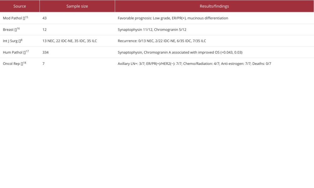
Table 2
https://jours.isi-science.com/imageXml.php?i=t2-amjcaserep-26-e950684.jpg&idArt=950684&w=1000
Table 2
Bảng 2
Additional studies.
Các nghiên cứu bổ sung.
Có thể bạn quan tâm
-
Viêm mô tế bào tái phát không sốt biến chứng do nhiễm toan lactic liên quan đến linezolid ở một bệnh nhân cao tuổi
Afebrile Recurrent Cellulitis Complicated by Linezolid-Induced Lactic Acidosis in an Elderly Patient
-
Thay thế bác sĩ bằng điều dưỡng trong môi trường bệnh viện: Hiệu quả đối với kết cục của bệnh nhân, quy trình chăm sóc và chi phí
Nurse-physician substitution in hospital settings: Effects on patient outcomes, care processes, and costs
-
Dung dịch đệm so với nước muối sinh học 0,9% trong hồi sức ở người lớn và trẻ em nguy kịch
Buffered solutions versus 0.9% saline for resuscitation in critically ill adults and children
-
Bác sĩ được đào tạo ở nước ngoài duy trì hệ thống y tế NZ – chúng tôi đã không phải lúc nào cũng chào đón như vậy
Foreign-trained doctors sustain NZ’s health system – we weren’t always so welcoming