Ca lâm sàng
Thách thức trong chẩn đoán các khối u vùng bụng không có đặc điểm hình ảnh điển hình: Từ chẩn đoán nhầm u tuyến nang tụy đến u tủy thượng thận và phản ứng xử trí trong mổ
Diagnostic Challenges of Abdominal Tumors Without Typical Imaging Features: From Misdiagnosis of Pancreatic Cystadenoma to Adrenal Pheochromocytoma and the Intraoperative Response
Haoyang Huang; Dingwei Xu; Zhangbin Chen; Jie Huang. American Journal of Case Reports. doi: 10.12659/AJCR.950574. Open access.
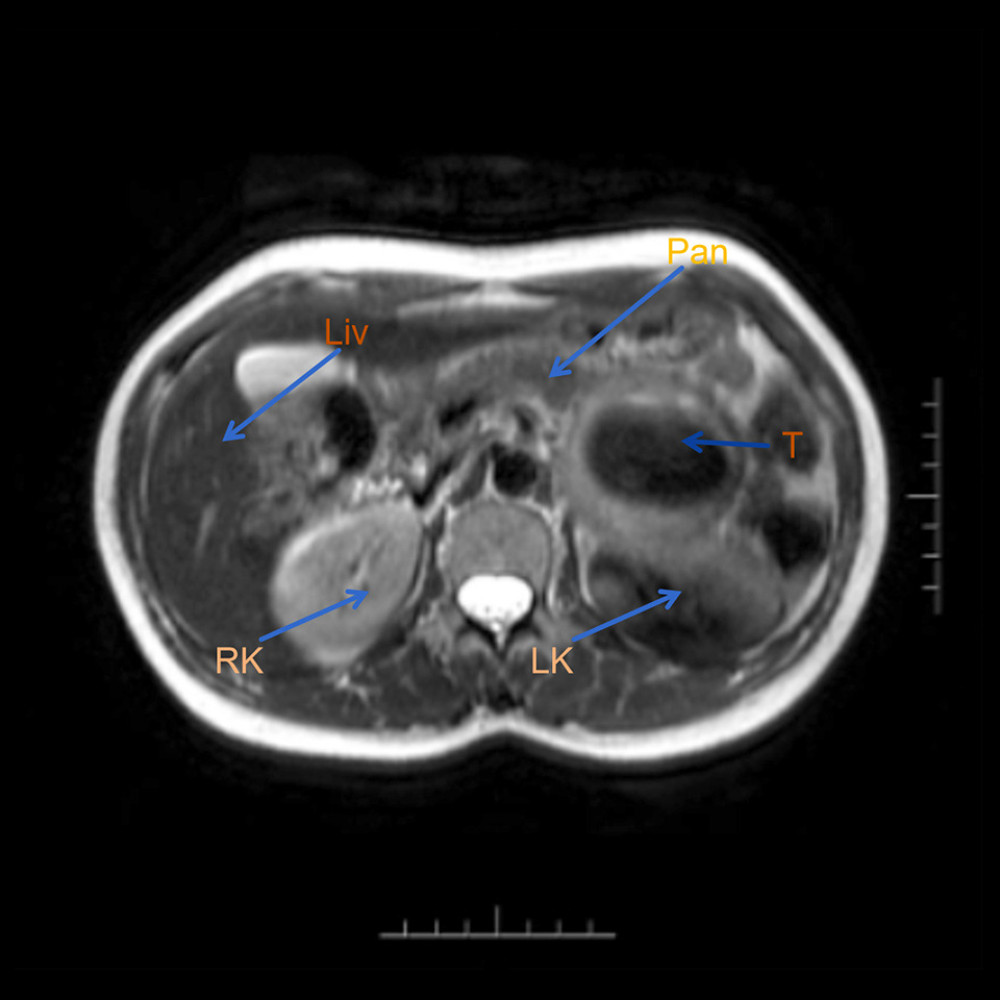
Figure 1
https://jours.isi-science.com/imageXml.php?i=amjcaserep-26-e950574-g001.jpg&idArt=950574&w=1000
Figure 1
Hình 1
Preoperative MRI (RK – right kidney; LK – left kidney; T – tumor; Pan – pancreas; Liv – liver).
Phim chụp cộng hưởng từ (MRI) trước mổ (RK – thận phải; LK – thận trái; T – khối u; Pan – tuyến tụy; Liv – gan).
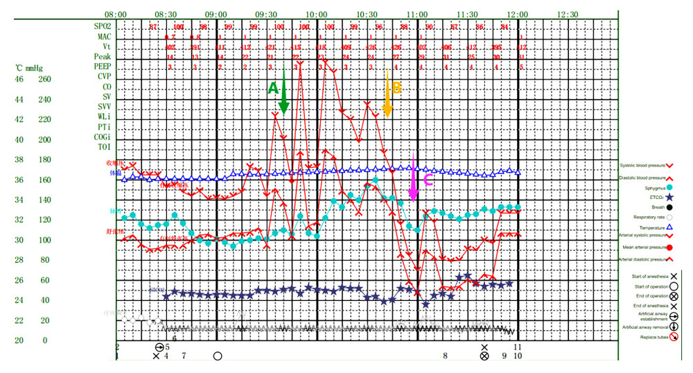
Figure 2
https://jours.isi-science.com/imageXml.php?i=amjcaserep-26-e950574-g002.jpg&idArt=950574&w=1000
Figure 2
Hình 2
Intraoperative anesthesia record. A) Use sodium nitroprusside. B) Use hydrocortisone. C) Use norepinephrine and metaraminol.
Bản ghi gây mê trong mổ. A) Sử dụng natri nitroprusside. B) Sử dụng hydrocortisone. C) Sử dụng norepinephrine và metaraminol.
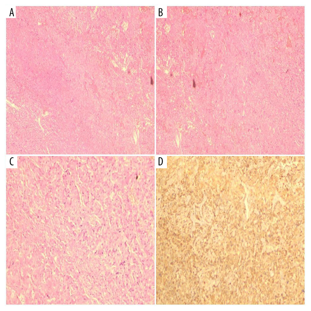
Figure 3
https://jours.isi-science.com/imageXml.php?i=amjcaserep-26-e950574-g003.jpg&idArt=950574&w=1000
Figure 3
Hình 3
Pathology results showing adrenal pheochromocytoma. (A, B) Staining with hematoxylin-eosin (HE); Observation perspective: Two-dimensional planar observation; Internal scale: 1 unit=100–200 μm (low-power). (C) Staining with hematoxylin-eosin (HE); Observation perspective: Two-dimensional planar observation; Internal scale: 1 unit=25–50 μm (high-power). (D) Immunohistochemical staining; Observation perspective: Two-dimensional planar observation; Internal scale: 1 unit=25–50 μm (high-power); Immunohistochemical Results: S100(+), PMS-2 (machine-stained) (+), MLH1 (machine-stained) (focal +), MSH2 (machine-stained) (focal +), MSH6 (machine-stained) (+), β-catenin (membranous +), inhibin-α(−), Ki-67(1%), ATRX(+), SF-1 (+), P27(+), CD56(+), CgA(+), Syn(+), CKL(−), SDHB(+).
Kết quả giải phẫu bệnh cho thấy u tủy thượng thận. (A, B) Nhuộm hematoxylin-eosin (HE); Góc độ quan sát: Quan sát mặt phẳng hai chiều; Thước đo bên trong: 1 đơn vị = 100–200 μm (vật kính nhỏ). (C) Nhuộm hematoxylin-eosin (HE); Góc độ quan sát: Quan sát mặt phẳng hai chiều; Thước đo bên trong: 1 đơn vị = 25–50 μm (vật kính lớn). (D) Nhuộm hóa mô miễn dịch; Góc độ quan sát: Quan sát mặt phẳng hai chiều; Thước đo bên trong: 1 đơn vị = 25–50 μm (vật kính lớn); Kết quả hóa mô miễn dịch: S100(+), PMS-2 (nhuộm máy) (+), MLH1 (nhuộm máy) (+ khu trú), MSH2 (nhuộm máy) (+ khu trú), MSH6 (nhuộm máy) (+), β-catenin (+ ở màng tế bào), inhibin-α(−), Ki-67(1%), ATRX(+), SF-1 (+), P27(+), CD56(+), CgA(+), Syn(+), CKL(−), SDHB(+).
Có thể bạn quan tâm
-
Mang thai sau khi hóa trị điều trị ung thư biểu mô màng đệm trong nhau thai: Theo dõi 5 năm thuận lợi đối với mẹ và con
Conception After Chemotherapy for Intraplacental Choriocarcinoma: Favorable 5-Year Follow-Up of the Mother and Child
-
Hiệu quả của các can thiệp kích thích hoặc phản hồi sinh học trong kiểm soát tình trạng mệt mỏi sau chấn thương sọ não (TBI)
Effects of stimulation or biofeedback interventions for fatigue management after traumatic brain injury (TBI)
-
Các chương trình đào tạo hồi sức chính quy chuẩn hóa nhằm giảm tỷ lệ tử vong và tỷ lệ mắc bệnh ở trẻ sơ sinh
Standardised formal resuscitation training programmes for reducing mortality and morbidity in newborn infants
-
Song ngữ và hormone giới tính có thể cung cấp một mối liên hệ mới với khả năng phục hồi của não và nguy cơ sa sút trí tuệ
Bilingualism and sex hormones may provide a new link to brain resilience and dementia risk