Ca lâm sàng
Hội chứng khó thở – giảm oxy máu khi đứng liên quan đến luồng thông tim phải-sang-trái được phát hiện bằng xạ hình tưới máu phổi
Platypnea-Orthodeoxia Syndrome Associated With a Right-to-Left Cardiac Shunt Detected by Lung Perfusion Scintigraphy
Yusuke Nakano; Hirohiko Ando; Akinori Satake; Reiji Goto; Kentaro Mukai; Tetsuya Amano. American Journal of Case Reports. doi: 10.12659/AJCR.949642. Open access.
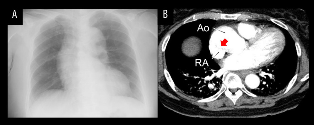
Figure 1
https://jours.isi-science.com/imageXml.php?i=amjcaserep-26-e949642-g001.jpg&idArt=949642&w=1000
Figure 1
Hình 1
Chest imaging findings. (A) Chest X-ray showing mild cardiomegaly and dilation of the right pulmonary hilum. (B) Contrast-enhanced chest CT scan demonstrating compression of the right atrium by an enlarged ascending aorta (red arrow). CT – computed tomography; Ao – aorta; RA – right atrium.
Các phát hiện trên hình ảnh học ngực. (A) X-quang ngực cho thấy bóng tim to nhẹ và giãn rốn phổi phải. (B) Phim chụp CT ngực có cản quang cho thấy tâm nhĩ phải bị chèn ép bởi động mạch chủ lên bị giãn rộng (mũi tên đỏ). CT – chụp cắt lớp vi tính; Ao – động mạch chủ; RA – tâm nhĩ phải.
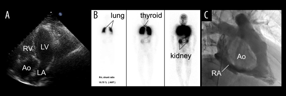
Figure 2
https://jours.isi-science.com/imageXml.php?i=amjcaserep-26-e949642-g002.jpg&idArt=949642&w=1000
Figure 2
Hình 2
Cardiac and scintigraphic findings. (A) Apical 4-chamber view on transthoracic echocardiography showing an enlarged aortic root (Ao) obscuring visualization of the right atrium, tricuspid valve, and interatrial septum, where a shunt would usually be identified. (B) Lung perfusion scintigraphy demonstrating technetium-99m (99mTc) particle uptake in the thyroid (middle) and kidneys (right) in addition to the lungs (left), indicating systemic distribution due to a right-to-left shunt. (C) Right atriography showing compression of the right atrium by the dilated ascending aorta (Ao). 99mTc – technetium-99m-labeled macroaggregated albumin; Ao – aorta; RV – right ventricle; LV – left ventricle; LA – left atrium; RA – right atrium.
Các phát hiện về tim và xạ hình. (A) Mặt cắt 4 buồng từ mỏm trên siêu âm tim qua thành ngực cho thấy gốc động mạch chủ (Ao) bị giãn rộng làm mờ hình ảnh quan sát tâm nhĩ phải, van ba lá và vách liên nhĩ, nơi thường xác định luồng thông. (B) Xạ hình tưới máu phổi cho thấy sự hấp thu hạt technetium-99m (99mTc) ở tuyến giáp (giữa) và thận (phải) bên cạnh phổi (trái), biểu hiện sự phân bố vào hệ tuần hoàn hệ thống do luồng thông phải-sang-trái. (C) Chụp buồng tâm nhĩ phải cho thấy tâm nhĩ phải bị chèn ép bởi động mạch chủ lên bị giãn (Ao). 99mTc – macroaggregated albumin gắn nhãn technetium-99m; Ao – động mạch chủ; RV – tâm thất phải; LV – tâm thất trái; LA – tâm nhĩ trái; RA – tâm nhĩ phải.
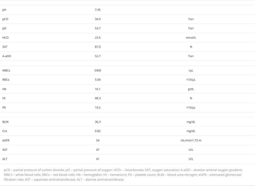
Table 1
https://jours.isi-science.com/imageXml.php?i=t1-amjcaserep-26-e949642.jpg&idArt=949642&w=1000
Table 1
Bảng 1
Admission laboratory data: supine hypoxemia with mild hepatic dysfunction and polycythemia.
Dữ liệu xét nghiệm lúc nhập viện: tình trạng giảm oxy máu khi nằm ngửa kèm theo rối loạn chức năng gan nhẹ và đa hồng cầu.
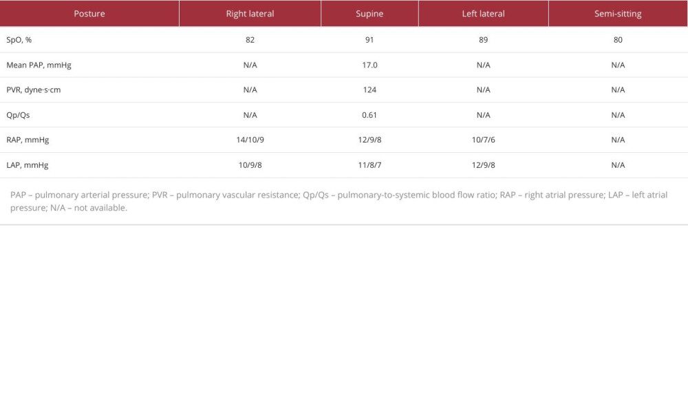
Table 2
https://jours.isi-science.com/imageXml.php?i=t2-amjcaserep-26-e949642.jpg&idArt=949642&w=1000
Table 2
Bảng 2
Oxygen saturation and right and left atrial pressures in various postures.
Độ bão hòa oxy cùng áp lực tâm nhĩ phải và trái ở các tư thế khác nhau.
Có thể bạn quan tâm
-
Đáp ứng với hormone tăng trưởng ở trẻ có đột biến TOMM7 đồng hợp tử: Những góc nhìn điều trị mới
Growth Hormone Response in a Child With a Homozygous TOMM7 Mutation: Novel Therapeutic Insights
-
Ảnh hưởng của các phương pháp phẫu thuật sa tạng chậu (POP) khác nhau, có và không có các thủ thuật giữ tiểu đồng thời, đối với phụ nữ mắc són tiểu khi gắng sức (SUI) và sa tạng chậu (POP)
Effects of different POP surgical approaches, with and without concomitant continence procedures, for women with SUI and POP
-
Hiệu quả và tính an toàn của VPM1002 và Immuvac trong phòng ngừa bệnh lao: thử nghiệm lâm sàng ngẫu nhiên pha 3 (Thử nghiệm PreVenTB)
Efficacy and safety of VPM1002 and Immuvac in preventing tuberculosis: phase 3 randomised clinical trial (PreVenTB trial)
-
Khủng hoảng thai chết lưu tiềm ẩn ở châu Phi: Báo cáo mới vạch trần những lỗ hổng lớn về chính sách và dữ liệu
Africa’s hidden stillbirth crisis: new report exposes major policy and data gaps