Bệnh giảm gammaglobulin máu Bruton: Chuỗi ca bệnh và tổng quan tài liệu từ Bệnh viện Trung ương King Fahad, Jizan, Ả Rập Xê Út

Bruton’s Agammaglobulinemia: Case Series and Literature Review From King Fahad Central Hospital, Jizan, Saudi Arabia

Ahmed Shamakhi; Nabil Dhayhi; Shatha Matabi; Manal Maashi; Abdullah Dhaifallah Hamdi; Bander Ali; Fadhel Hazazi; Abdullah H. Alhamoud. American Journal of Case Reports. doi: 10.12659/AJCR.949936. Open access.
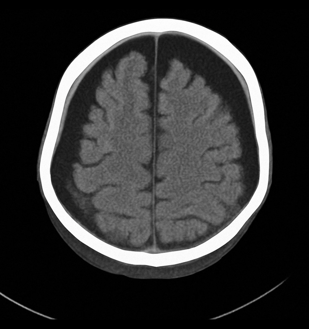
Axial computed tomography scan of the brain showing bilateral subdural hygroma and mild cerebral atrophy, consistent with chronic intracranial complications secondary to recurrent infections in X-linked agammaglobulinemia (Case 1).
Figure 1
https://jours.isi-science.com/imageXml.php?i=amjcaserep-26-e949936-g001.jpg&idArt=949936&w=1000 Figure 1 Hình 1
Axial computed tomography scan of the brain showing bilateral subdural hygroma and mild cerebral atrophy, consistent with chronic intracranial complications secondary to recurrent infections in X-linked agammaglobulinemia (Case 1).
Hình ảnh chụp cắt lớp vi tính sọ não cắt ngang (axial) cho thấy khối tụ dịch dưới màng cứng hai bên và teo não nhẹ, phù hợp với các biến chứng nội sọ mạn tính thứ phát sau các đợt nhiễm trùng tái phát trong bệnh giảm gammaglobulin máu liên kết nhiễm sắc thể X (Ca bệnh 1).
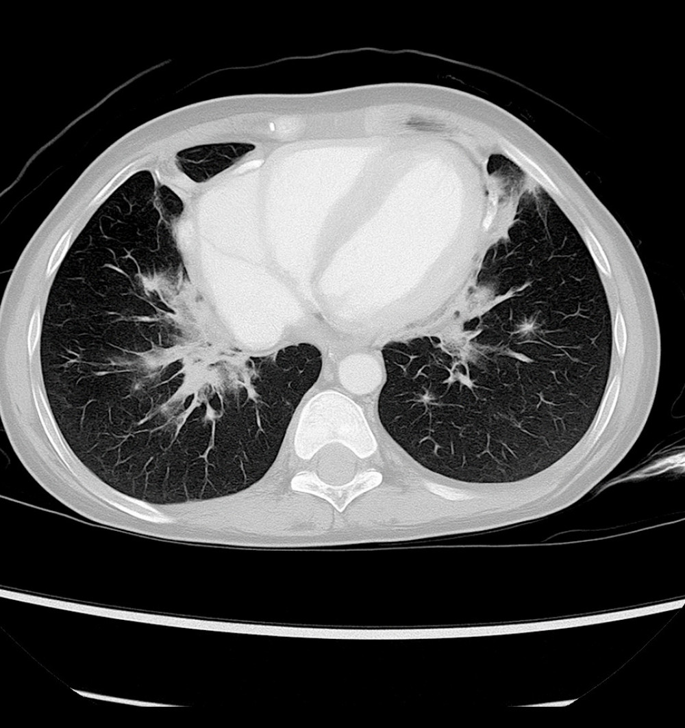
Axial chest computed tomography image showing bilateral pulmonary infiltrates with areas of patchy consolidation (Case 2).
Figure 2
https://jours.isi-science.com/imageXml.php?i=amjcaserep-26-e949936-g002.jpg&idArt=949936&w=1000 Figure 2 Hình 2
Axial chest computed tomography image showing bilateral pulmonary infiltrates with areas of patchy consolidation (Case 2).
Hình ảnh chụp cắt lớp vi tính lồng ngực cắt ngang (axial) cho thấy các tổn thương thâm nhiễm phổi hai bên kèm theo các vùng đông đặc rải rác thành đám (Ca bệnh 2).
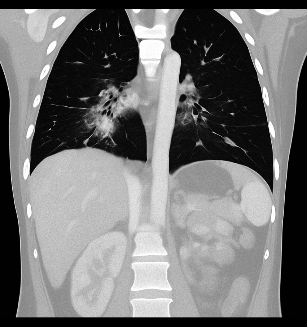
Coronal chest computed tomography image showing ground-glass opacities predominantly in the perihilar regions, consistent with an infectious or inflammatory process (Case 2).
Figure 3
https://jours.isi-science.com/imageXml.php?i=amjcaserep-26-e949936-g003.jpg&idArt=949936&w=1000 Figure 3 Hình 3
Coronal chest computed tomography image showing ground-glass opacities predominantly in the perihilar regions, consistent with an infectious or inflammatory process (Case 2).
Hình ảnh chụp cắt lớp vi tính lồng ngực đứng dọc (coronal) cho thấy các vùng kính mờ chiếm ưu thế ở vùng quanh rốn phổi, phù hợp với diễn tiến nhiễm trùng hoặc viêm (Ca bệnh 2).
Show
Hide
EN – VI
VI – EN

Abstract

Tóm tắt

BACKGROUND: X-linked agammaglobulinemia (XLA), also known as Bruton’s agammaglobulinemia, is a rare genetic disorder caused by mutations in the Bruton tyrosine kinase (BTK) gene. This defect leads to a substantial reduction in B cells and immunoglobulins, predisposing patients to recurrent severe infections. Early diagnosis and regular immunoglobulin replacement therapy are essential for effective disease management.

BỐI CẢNH: Bệnh giảm gammaglobulin máu liên kết nhiễm sắc thể X (XLA), hay còn gọi là bệnh giảm gammaglobulin máu Bruton, là một rối loạn di truyền hiếm gặp do các đột biến ở gen Bruton tyrosine kinase (BTK) gây ra. Khiếm khuyết này dẫn đến sự sụt giảm nghiêm trọng các tế bào B và globulin miễn dịch, khiến bệnh nhân dễ bị nhiễm trùng nặng tái phát. Chẩn đoán sớm và liệu pháp thay thế globulin miễn dịch thường xuyên là những yếu tố thiết yếu để quản lý bệnh hiệu quả.

CASE REPORT: This report describes 2 male pediatric patients diagnosed with XLA at King Fahad Central Hospital in Jizan, Saudi Arabia. The first case involved a 14-month-old boy with recurrent respiratory infections, developmental delay, and frequent hospital admissions. Despite receiving immunoglobulin replacement therapy, his condition deteriorated due to poor treatment adherence, ultimately resulting in death. The second case involved a 3-year-old boy with recurrent respiratory infections and severe neutropenia. Genetic testing confirmed a BTK gene mutation. Consistent intravenous immunoglobulin therapy and close monitoring improved infection control and clinical stability, although he remained at risk for infections.

BÁO CÁO CA BỆNH: Báo cáo này mô tả 2 bệnh nhi nam được chẩn đoán mắc XLA tại Bệnh viện Trung ương King Fahad ở Jizan, Ả Rập Xê Út. Ca bệnh đầu tiên liên quan đến một bé trai 14 tháng tuổi bị nhiễm trùng hô hấp tái phát, chậm phát triển và thường xuyên phải nhập viện. Mặc dù được điều trị bằng liệu pháp thay thế globulin miễn dịch, tình trạng của bệnh nhi vẫn chuyển biến xấu do tuân thủ điều trị kém, cuối cùng dẫn đến tử vong. Ca bệnh thứ hai liên quan đến một bé trai 3 tuổi bị nhiễm trùng hô hấp tái phát và giảm bạch cầu trung tính nghiêm trọng. Xét nghiệm di truyền đã xác nhận có đột biến gen BTK. Liệu pháp tiêm tĩnh mạch globulin miễn dịch liên tục kết hợp theo dõi chặt chẽ đã giúp cải thiện khả năng kiểm soát nhiễm trùng và độ ổn định lâm sàng, mặc dù bệnh nhi vẫn có nguy cơ bị nhiễm trùng.

CONCLUSIONS: These cases emphasize the importance of early genetic diagnosis and strict adherence to immunoglobulin therapy in managing XLA. The differing outcomes underscore the critical role of treatment compliance in preventing severe complications and improving long-term prognosis. In regions with high consanguinity, genetic testing and family counseling are vital for early detection and optimal disease management.

KẾT LUẬN: Các ca bệnh này nhấn mạnh tầm quan trọng của việc chẩn đoán di truyền sớm và tuân thủ nghiêm ngặt liệu pháp globulin miễn dịch trong quản lý XLA. Kết quả khác nhau giữa hai ca bệnh khẳng định vai trò quyết định của việc tuân thủ điều trị trong việc ngăn ngừa các biến chứng nghiêm trọng và cải thiện tiên lượng lâu dài. Ở những vùng có tỷ lệ kết hôn cận huyết cao, xét nghiệm di truyền và tư vấn gia đình là những mắt xích quan trọng để phát hiện sớm và quản lý bệnh tối ưu.

Keywords: agammaglobulinemia, immunocompromised host, infections

Từ khóa: giảm gammaglobulin máu, ký chủ suy giảm miễn dịch, nhiễm trùng

Introduction

Đặt vấn đề

X-linked agammaglobulinemia (XLA), also known as Bruton’s agammaglobulinemia, is a primary immunodeficiency affecting the humoral immune response, first described by Bruton in 1952 1. The disorder results from mutations in the Bruton tyrosine kinase (BTK) gene, located on the Xq22 region of the X chromosome, which is essential for normal B-cell development 1,2. Mutations in the BTK gene disrupt B-cell maturation, resulting in substantially reduced levels of mature B lymphocytes, plasma cells, and various immunoglobulin (Ig) isotypes 3. Consequently, patients with XLA are highly susceptible to recurrent infections, particularly respiratory infections caused by encapsulated bacteria, and gastrointestinal infections due to pathogens such as Salmonella and Giardia 4. The cornerstone of XLA management is immunoglobulin replacement therapy, administered either intravenously (IVIG) or subcutaneously 5.

Bệnh giảm gammaglobulin máu liên kết nhiễm sắc thể X (XLA), hay còn gọi là bệnh giảm gammaglobulin máu Bruton, là một tình trạng suy giảm miễn dịch bẩm sinh ảnh hưởng đến đáp ứng miễn dịch dịch thể, được Bruton mô tả lần đầu tiên vào năm 1952 1. Rối loạn này là hệ quả của các đột biến ở gen Bruton tyrosine kinase (BTK), nằm trên vùng Xq22 của nhiễm sắc thể X, vốn đóng vai trò thiết yếu cho sự phát triển bình thường của tế bào B 1,2. Các đột biến ở gen BTK làm gián đoạn quá trình trưởng thành của tế bào B, dẫn đến sự sụt giảm nghiêm trọng nồng độ của các lympho bào B trưởng thành, tương bào và các kiểu đồng hình globulin miễn dịch (Ig) khác nhau 3. Hệ quả là bệnh nhân mắc XLA rất dễ bị nhiễm trùng tái phát, đặc biệt là nhiễm trùng đường hô hấp do vi khuẩn có vỏ bọc và nhiễm trùng đường tiêu hóa do các tác nhân như Salmonella và Giardia 4. Nền tảng trong quản lý XLA là liệu pháp thay thế globulin miễn dịch, được dùng thông qua đường tiêm tĩnh mạch (IVIG) hoặc tiêm dưới da 5.

The prevalence of XLA varies by region, with approximately 250 cases per 100,000 men reported in Saudi Arabia and around 1 case per 190,000 men in the United States 6,7. Diagnosis typically involves measuring serum immunoglobulin concentrations, enumerating B cells, and performing genetic testing to identify BTK mutations. More than 600 pathogenic variants of the BTK gene have been documented, and no single variant is predominant 8–11. These genetic differences contribute to the heterogeneity of XLA, which may manifest with atypical features, including both IgE-mediated and non-IgE-mediated allergic reactions 12–14. Given the high consanguinity rates in Saudi Arabia – ranging from 44% to 67% nationally and reaching 66.7% in the Jazan region – the likelihood of inherited immunodeficiency syndromes such as XLA is elevated in this population 15.

Tỷ lệ lưu hành của XLA có sự khác biệt theo từng khu vực, với khoảng 250 ca trên 100.000 nam giới được báo cáo ở Ả Rập Xê Út và khoảng 1 ca trên 190.000 nam giới ở Hoa Kỳ 6,7. Việc chẩn đoán thường bao gồm định lượng nồng độ globulin miễn dịch trong huyết thanh, đếm số lượng tế bào B và thực hiện xét nghiệm di truyền để xác định các đột biến BTK. Cho đến nay, hơn 600 biến thể gây bệnh của gen BTK đã được ghi nhận và không có biến thể đơn lẻ nào chiếm ưu thế 8–11. Những khác biệt về mặt di truyền này góp phần tạo nên tính không đồng nhất của XLA, bệnh có thể biểu hiện bằng các đặc điểm không điển hình, bao gồm cả các phản ứng dị ứng trung gian IgE và không trung gian IgE 12–14. Do tỷ lệ kết hôn cận huyết cao ở Ả Rập Xê Út – dao động từ 44% đến 67% trên toàn quốc và đạt mức 66,7% ở vùng Jazan – khả năng mắc các hội chứng suy giảm miễn dịch di truyền như XLA được ghi nhận là tăng cao ở quần thể dân cư này 15.

This case series and literature review aim to describe the clinical and genetic characteristics of XLA observed at King Fahad Central Hospital in Jizan, Saudi Arabia, thereby contributing valuable data to the global understanding of this rare and serious immunodeficiency disorder.

Chuỗi ca bệnh và tổng quan tài liệu này nhằm mục đích mô tả các đặc điểm lâm sàng và di truyền của XLA được quan sát tại Bệnh viện Trung ương King Fahad ở Jizan, Ả Rập Xê Út, từ đó đóng góp những dữ liệu có giá trị vào sự hiểu biết toàn cầu về rối loạn suy giảm miễn dịch hiếm gặp và nghiêm trọng này.

Case Reports

Báo cáo ca bệnh

CASE 1:

CA BỆNH 1:

A 14-month-old male child had a history of recurrent chest infections and significant developmental delay; eventually, he was diagnosed with agammaglobulinemia via whole-exome sequencing. The analysis identified a hemizygous pathogenic BTK variant (c.953_956del; p.S318Cfs*12) resulting in a frameshift and premature protein truncation, thus confirming the diagnosis of XLA. The patient had been born at home through an uncomplicated vaginal delivery without prenatal care. His early development was normal until approximately 5 months of age, when he began to exhibit developmental regression and frequent respiratory infections, leading to multiple hospital admissions.

Một bé trai 14 tháng tuổi có tiền sử nhiễm trùng ngực tái phát và chậm phát triển nghiêm trọng; cuối cùng, bệnh nhi được chẩn đoán mắc bệnh giảm gammaglobulin máu thông qua giải trình tự toàn bộ vùng mã hóa (whole-exome sequencing). Phân tích đã xác định một biến thể BTK gây bệnh ở trạng thái dị hợp tử tử (c.953_956del; p.S318Cfs*12) dẫn đến dịch khung và cắt cụt protein sớm, qua đó xác nhận chẩn đoán mắc XLA. Bệnh nhi được sinh tại nhà bằng phương pháp sinh thường qua đường âm đạo không có biến chứng và không được chăm sóc tiền sản. Sự phát triển sớm của trẻ bình thường cho đến khoảng 5 tháng tuổi, khi trẻ bắt đầu có dấu hiệu thoái lui phát triển và nhiễm trùng hô hấp thường xuyên, dẫn đến nhiều lần phải nhập viện.

By 14 months of age, his condition had worsened. He was referred to a peripheral hospital after 6 days of fever, cough, and diarrhea. Despite treatment with antibiotics and antiviral agents, including oseltamivir, cefotaxime, and vancomycin, his clinical condition remained unstable. Eventually, he was hospitalized more than 5 times for respiratory infections, raising suspicion of agammaglobulinemia due to the infection frequency and severity.

Đến 14 tháng tuổi, tình trạng của bệnh nhi trở nên tồi tệ hơn. Trẻ được chuyển đến một bệnh viện tuyến dưới sau 6 ngày bị sốt, ho và tiêu chảy. Mặc dù được điều trị bằng các thuốc kháng sinh và kháng virus, bao gồm oseltamivir, cefotaxime và vancomycin, tình trạng lâm sàng của trẻ vẫn không ổn định. Cuối cùng, trẻ đã phải nhập viện hơn 5 lần vì nhiễm trùng hô hấp, làm dấy lên nghi ngờ mắc bệnh giảm gammaglobulin máu do tần suất và mức độ nghiêm trọng của các đợt nhiễm trùng.

Physical examination revealed edema of the lower limbs, hands, and periorbital regions, along with coarse facial features. His abdomen was distended; he exhibited cutaneous lesions and reduced air entry in the lower left lung. Laboratory investigations demonstrated severe immunodeficiency, with greatly decreased levels of all immunoglobulin classes: IgM levels ranged from 0.03 to 0.15 g/L (reference: 0.4–2.3 g/L), IgG levels from 0.03 to 13.55 g/L (reference: 7–16 g/L), and IgA levels from 0 to 0.08 g/L (reference: 0.7–4 g/L). Complete blood count results showed clinically significant abnormalities, including white blood cell counts fluctuating from 2.14 × 10⁹/L to 18.64×10⁹/L (reference: 4.5–10×109/L), persistently low hemoglobin levels around 6.3 g/dL (reference: 13–18 g/dL), and platelet counts varying from 12 to 563×109/L (reference: 150–400×109/L). He also had recurrent episodes of severe neutropenia, with neutrophil counts as low as 0.3×10⁹/L (reference: 2.5–7.5×10⁹/L), and elevated lymphocyte counts up to 13.69×10⁹/L (reference: 1.5–3.5×10⁹/L), further increasing his susceptibility to infections.

Thăm khám lâm sàng ghi nhận tình trạng phù nề ở các chi dưới, bàn tay và vùng quanh ổ mắt, cùng với các đường nét khuôn mặt thô. Bụng của trẻ bị chướng; trẻ có các tổn thương da và giảm thông khí ở phổi dưới bên trái. Các xét nghiệm cận lâm sàng cho thấy tình trạng suy giảm miễn dịch nghiêm trọng, với nồng độ của tất cả các nhóm globulin miễn dịch giảm mạnh: nồng độ IgM dao động từ 0,03 đến 0,15 g/L (khoảng tham chiếu: 0,4–2,3 g/L), nồng độ IgG từ 0,03 đến 13,55 g/L (khoảng tham chiếu: 7–16 g/L) và nồng độ IgA từ 0 đến 0,08 g/L (khoảng tham chiếu: 0,7–4 g/L). Kết quả tổng phân tích tế bào máu cho thấy những bất thường có ý nghĩa lâm sàng, bao gồm số lượng bạch cầu dao động từ 2,14 × 10⁹/L đến 18,64×10⁹/L (khoảng tham chiếu: 4,5–10×109/L), nồng độ hemoglobin thấp kéo dài khoảng 6,3 g/dL (khoảng tham chiếu: 13–18 g/dL) và số lượng tiểu cầu thay đổi từ 12 đến 563×109/L (khoảng tham chiếu: 150–400×109/L). Bệnh nhi cũng có các đợt giảm bạch cầu trung tính nghiêm trọng tái diễn, với số lượng bạch cầu trung tính thấp tới mức 0,3×10⁹/L (khoảng tham chiếu: 2,5–7.5×10⁹/L), và số lượng lympho bào tăng cao lên tới 13,69×10⁹/L (khoảng tham chiếu: 1,5–3,5×10⁹/L), càng làm tăng thêm khả năng dễ bị nhiễm trùng.

Imaging studies, including a computed tomography (CT) scan of the brain, revealed a subdural hygroma and mild cerebral atrophy (Figure 1). As the child grew older, adherence to the recommended monthly IVIG treatments declined, worsening his condition. At 2 years of age, he was admitted to the hospital with severe respiratory distress after missing 3 consecutive months of IVIG therapy due to social difficulties. He presented with fever, shortness of breath, and a worsening cough.

Các thăm dò chẩn đoán hình ảnh, bao gồm chụp cắt lớp vi tính (CT) não, phát hiện một khối tụ dịch dưới màng cứng và teo não nhẹ (Hình 1). Khi trẻ lớn hơn, việc tuân thủ các liệu pháp điều trị IVIG hàng tháng theo khuyến cáo bị giảm sút, khiến tình trạng của trẻ ngày càng trầm trọng. Vào năm 2 tuổi, bệnh nhi phải nhập viện vì suy hô hấp nghiêm trọng sau khi bỏ lỡ liệu pháp IVIG trong 3 tháng liên tiếp do hoàn cảnh xã hội khó khăn. Trẻ nhập viện với các triệu chứng sốt, khó thở và ho nặng hơn.

On examination, the patient was tachypneic (130 breaths/min), in respiratory distress, and exhibiting a distended abdomen with non-pitting edema of the lower limbs. Chest auscultation revealed bilateral wheezing and crackles. Despite aggressive management, including high-flow nasal cannula oxygen therapy, IVIG administration, and broad-spectrum antibiotics such as meropenem and vancomycin, his condition continued to deteriorate.

Khi thăm khám, bệnh nhân có biểu hiện thở nhanh (130 lần/phút), suy hô hấp và bụng chướng kèm theo phù mềm không ấn lõm ở các chi dưới. Nghe phổi phát hiện có tiếng rít và răng ẩm cả hai bên. Mặc dù được xử trí tích cực, bao gồm liệu pháp oxy qua canula mũi dòng cao, truyền IVIG và sử dụng các kháng sinh phổ rộng như meropenem và vancomycin, tình trạng của trẻ vẫn tiếp tục chuyển biến xấu.

Throughout his illness, the patient experienced recurrent episodes of severe respiratory distress, frequently warranting intensive care. He was treated for multiple infections, including pneumonia, and required management of complications related to his immunodeficiency. Despite repeated interventions and the administration of IVIG, his prognosis remained poor. His clinical course was further complicated by inconsistent adherence to therapy, resulting in recurrent infections and repeated hospitalizations. Ultimately, the patient succumbed to his illness, primarily due to severe and recurrent respiratory infections associated with XLA and irregular treatment adherence.

Trong suốt quá trình bệnh, bệnh nhân phải trải qua các đợt suy hô hấp nghiêm trọng tái diễn, thường xuyên cần đến sự chăm sóc tích cực. Trẻ đã được điều trị nhiều đợt nhiễm trùng, bao gồm cả viêm phổi, và cần phải quản lý các biến chứng liên quan đến tình trạng suy giảm miễn dịch của mình. Mặc dù đã có nhiều can thiệp lặp lại và tiến hành truyền IVIG, tiên lượng của trẻ vẫn rất kém. Diễn tiến lâm sàng của bệnh nhi càng trở nên phức tạp do việc tuân thủ điều trị không liên tục, dẫn đến nhiễm trùng tái phát và phải nhập viện nhiều lần. Cuối cùng, bệnh nhân đã tử vong do bệnh tật, nguyên nhân chủ yếu là do các đợt nhiễm trùng hô hấp nghiêm trọng và tái diễn liên quan đến XLA kết hợp với việc tuân thủ điều trị không đều đặn.

The patient’s parents provided informed consent, verbally agreeing that the patient’s data could be used for this publication.

Cha mẹ của bệnh nhân đã cung cấp văn bản đồng thuận, đồng ý bằng lời rằng dữ liệu của bệnh nhân có thể được sử dụng cho bài công bố này.

CASE 2:

CA BỆNH 2:

A 3-year-old male child experienced repeated respiratory infections and severe neutropenia, leading to an eventual diagnosis of XLA. His history of repeated chest infections began at approximately 5 months of age, raising early suspicion of an underlying immunodeficiency. His first hospitalization was prompted by symptoms of gastroenteritis, including diarrhea and decreased oral intake, accompanied by severe neutropenia. Although he remained hydrated and alert, imaging studies (including CT scans) revealed bilateral pulmonary infiltrates with areas of consolidation (Figure 2) and ground-glass opacities consistent with pneumonia (Figure 3).

Một bé trai 3 tuổi gặp phải các đợt nhiễm trùng hô hấp lặp đi lặp lại và giảm bạch cầu trung tính nghiêm trọng, cuối cùng dẫn đến chẩn đoán mắc XLA. Tiền sử nhiễm trùng ngực nhiều lần của trẻ bắt đầu từ khoảng 5 tháng tuổi, làm dấy lên nghi ngờ sớm về tình trạng suy giảm miễn dịch tiềm ẩn. Đợt nhập viện đầu tiên của trẻ là do các triệu chứng của viêm dạ dày ruột, bao gồm tiêu chảy và giảm lượng ăn uống qua đường miệng, đi kèm với tình trạng giảm bạch cầu trung tính nghiêm trọng. Mặc dù trẻ vẫn giữ được nước và tỉnh táo, các thăm dò chẩn đoán hình ảnh (bao gồm chụp CT) đã phát hiện các tổn thương thâm nhiễm phổi cả hai bên kèm theo các vùng đông đặc (Hình 2) và các vùng kính mờ phù hợp với bệnh cảnh viêm phổi (Hình 3).

The diagnosis of agammaglobulinemia was confirmed through whole-exome sequencing, which identified a hemizygous BTK variant (c.1873del; p.Tyr625Thrfs*58) causing a frameshift and premature protein truncation. This genetic finding supported the diagnosis of XLA; it was consistent with the patient’s clinical presentation of recurrent infections and greatly reduced immunoglobulin levels. Laboratory investigations showed profoundly low immunoglobulin levels: IgG was 3.29 g/L (reference: 7–16 g/L), IgM ranged from 0.124 to 0.25 g/L (reference: 0.4–2.3 g/L), and IgA was undetectable at 0 g/L (reference: 0.7–4 g/L). Total IgE was within normal limits at 49.8 IU/mL (reference: <60 IU/mL). His complete blood count consistently revealed severe neutropenia involving neutrophil counts as low as 0, clinically significant anemia with hemoglobin levels decreasing to 8.8 g/dL (reference: 13–18 g/dL), and thrombocytosis, with platelet counts reaching 759×109/L (reference: 150–400×109/L). These findings reflected his heightened susceptibility to infections and the persistent challenges of disease management.

Chẩn đoán bệnh giảm gammaglobulin máu được xác nhận thông qua giải trình tự toàn bộ vùng mã hóa, kết quả phát hiện một biến thể BTK gây bệnh ở trạng thái dị hợp tử tử (c.1873del; p.Tyr625Thrfs*58) gây ra dịch khung và cắt cụt protein sớm. Phát hiện di truyền này đã củng cố cho chẩn đoán mắc XLA; kết quả này hoàn toàn phù hợp với biểu hiện lâm sàng của bệnh nhân về các đợt nhiễm trùng tái phát và nồng độ globulin miễn dịch giảm mạnh. Các xét nghiệm cận lâm sàng cho thấy nồng độ globulin miễn dịch thấp ở mức báo động: IgG là 3,29 g/L (khoảng tham chiếu: 7–16 g/L), IgM dao động từ 0,124 đến 0,25 g/L (khoảng tham chiếu: 0,4–2,3 g/L) và không thể phát hiện được IgA ở mức 0 g/L (khoảng tham chiếu: 0,7–4 g/L). Nồng độ IgE toàn phần nằm trong giới hạn bình thường ở mức 49,8 IU/mL (khoảng tham chiếu: <60 IU/mL). Kết quả tổng phân tích tế bào máu liên tục cho thấy tình trạng giảm bạch cầu trung tính nghiêm trọng với số lượng bạch cầu trung tính có lúc giảm về 0, tình trạng thiếu máu có ý nghĩa lâm sàng với nồng độ hemoglobin giảm xuống còn 8,8 g/dL (khoảng tham chiếu: 13–18 g/dL) và tình trạng tăng tiểu cầu, với số lượng tiểu cầu đạt mức 759×109/L (khoảng tham chiếu: 150–400×109/L). Những phát hiện này phản ánh khả năng dễ bị nhiễm trùng tăng cao của trẻ và những thách thức dai dẳng trong việc quản lý bệnh.

Throughout his clinical course, the patient experienced multiple severe infections, including nosocomial sepsis and urinary tract infections, requiring frequent hospitalizations and intensive treatment with broad-spectrum antibiotics such as vancomycin and tazocin. Despite regular IVIG therapy, difficulties in maintaining consistent adherence – particularly concerning timely IVIG administration – led to intervals of increased vulnerability to infection.

Trong suốt diễn tiến lâm sàng, bệnh nhân đã gặp phải nhiều đợt nhiễm trùng nặng, bao gồm nhiễm khuẩn huyết bệnh viện và nhiễm trùng đường tiết niệu, đòi hỏi phải nhập viện thường xuyên và điều trị tích cực bằng các kháng sinh phổ rộng như vancomycin và tazocin. Mặc dù được điều trị bằng liệu pháp IVIG định kỳ, những khó khăn trong việc duy trì sự tuân thủ điều trị nhất quán – đặc biệt là liên quan đến việc truyền IVIG đúng hạn – đã dẫn đến những khoảng thời gian làm tăng tính dễ bị tổn thương trước tình trạng nhiễm trùng.

As the patient grew older, his condition stabilized to some extent with consistent IVIG therapy, although he continued to experience occasional respiratory infections. By the age of 4 years and 8 months, he was generally stable during follow-up visits, reporting no new complaints and exhibiting stable vital signs. Chest examinations remained clear, and he was active and afebrile. However, his clinical course was often disrupted by episodes of decreased appetite and activity, typically coinciding with acute infections. Despite these challenges, his management plan – which included regular IVIG therapy and close monitoring for infections – helped reduce the frequency and severity of his symptoms.

Khi bệnh nhi lớn hơn, tình trạng của trẻ đã ổn định ở một mức độ nhất định nhờ liệu pháp IVIG liên tục, mặc dù trẻ vẫn thỉnh thoảng bị nhiễm trùng hô hấp. Vào thời điểm 4 tuổi 8 tháng, trẻ nhìn chung ở trạng thái ổn định trong các lần khám theo dõi, không có than phiền mới và các dấu hiệu sinh tồn ổn định. Thăm khám ngực cho kết quả rõ ràng, trẻ hoạt động tốt và không sốt. Tuy nhiên, diễn tiến lâm sàng của trẻ thường bị gián đoạn bởi các đợt giảm cảm giác thèm ăn và giảm hoạt động, thường trùng khớp với các đợt nhiễm trùng cấp tính. Bất chấp những thách thức này, kế hoạch quản lý bệnh của trẻ – bao gồm liệu pháp IVIG thường quy và theo dõi chặt chẽ các tình trạng nhiễm trùng – đã giúp giảm tần suất và mức độ nghiêm trọng của các triệu chứng.

Although the patient’s outlook remained cautiously optimistic, the chronic nature of his condition and the recurrent infections requiring ongoing medical intervention complicated his clinical course. His experience underscored the importance of strict adherence to treatment regimens and continuous monitoring to prevent severe complications associated with XLA.

Mặc dù triển vọng của bệnh nhân vẫn ở mức lạc quan một cách thận trọng, tính chất mạn tính của bệnh và các đợt nhiễm trùng tái phát đòi hỏi phải có sự can thiệp y tế liên tục đã làm cho diễn tiến lâm sàng của trẻ trở nên phức tạp. Trường hợp của trẻ đã nhấn mạnh tầm quan trọng của việc tuân thủ nghiêm ngặt các phác đồ điều trị và theo dõi liên tục để ngăn ngừa các biến chứng nghiêm trọng liên quan đến XLA.

The patient’s parents provided informed consent, verbally agreeing that his data could be included in this publication.

Cha mẹ của bệnh nhân đã cung cấp văn bản đồng thuận, đồng ý bằng lời rằng dữ liệu của trẻ có thể được đưa vào bài công bố này.

Discussion

Bàn luận

LIMITATIONS:

HẠN CHẾ:

This case series is limited by its single-center design, which may restrict the generalizability of its findings. The small sample size of 2 cases also limits the ability to draw broader conclusions about XLA. Future studies with larger, multicenter cohorts and prospective designs are warranted to better assess long-term outcomes and optimize treatment strategies, particularly in regions with high consanguinity rates.

Chuỗi ca bệnh này bị giới hạn bởi thiết kế nghiên cứu đơn trung tâm, điều này có thể làm hạn chế khả năng tổng quát hóa các phát hiện của nghiên cứu. Kích thước mẫu nhỏ chỉ gồm 2 ca bệnh cũng làm hạn chế khả năng đưa ra các kết luận rộng hơn về XLA. Các nghiên cứu trong tương lai với cỡ mẫu lớn hơn, thiết kế đa trung tâm và đoàn hệ tiến cứu là cần thiết để đánh giá tốt hơn kết cục lâu dài và tối ưu hóa các chiến lược điều trị, đặc biệt là ở những vùng có tỷ lệ kết hôn cận huyết cao.

Conclusions

Kết luận

XLA presents substantial clinical challenges, primarily due to its association with recurrent severe infections. The cases described in this report highlight the critical roles of early diagnosis, consistent immunoglobulin replacement therapy, and close clinical monitoring in effective disease management. The differing outcomes observed underscore the importance of treatment adherence in preventing severe complications and improving long-term prognosis. Moreover, the identification of BTK gene mutations is essential for confirming the diagnosis and guiding family counseling, especially in regions with high consanguinity rates, such as Saudi Arabia. Ultimately, these findings highlight the need for continuous patient education and strict adherence to treatment protocols to optimize outcomes for patients with XLA.

XLA đặt ra những thách thức lâm sàng to lớn, chủ yếu là do mối liên hệ của bệnh với các đợt nhiễm trùng nghiêm trọng tái diễn. Các ca bệnh được mô tả trong báo cáo này đã làm nổi bật vai trò quyết định của việc chẩn đoán sớm, liệu pháp thay thế globulin miễn dịch nhất quán và theo dõi lâm sàng chặt chẽ trong quản lý bệnh hiệu quả. Những kết cục khác nhau được quan sát thấy đã nhấn mạnh tầm quan trọng của việc tuân thủ điều trị trong việc ngăn ngừa các biến chứng nghiêm trọng và cải thiện tiên lượng lâu dài. Hơn nữa, việc xác định các đột biến gen BTK là cần thiết để xác nhận chẩn đoán và hướng dẫn tư vấn gia đình, đặc biệt là ở những vùng có tỷ lệ kết hôn cận huyết cao như Ả Rập Xê Út. Cuối cùng, những phát hiện này nêu bật nhu cầu phải giáo dục bệnh nhân liên tục và tuân thủ nghiêm ngặt các phác đồ điều trị để tối ưu hóa kết cục cho bệnh nhân mắc XLA.

Reference

  1. Vetrie D, Vořechovský I, Sideras P, The gene involved in X-linked agammaglobulinemia is a member of the Src family of protein-tyrosine kinases: J Immunol, 2012; 188; 2948-55
  2. Conley ME, Early defects in B-cell development: Curr Opin Allergy Clin Immunol, 2002; 2; 517-22
  3. Ochs HD, Smith CI, X-linked agammaglobulinemia: A clinical and molecular analysis: Medicine (Baltimore), 1996; 75; 287-99
  4. Lederman HM, Winkelstein JA, X-linked agammaglobulinemia: an analysis of 96 patients: Medicine (Baltimore), 1985; 64; 145-56
  5. Shillitoe BMJ, Gennery AR, An update on X-linked agammaglobulinemia: Clinical manifestations and management: Curr Opin Allergy Clin Immunol, 2019; 19; 571-77
  6. Al-Attas RA, Rahi AH, Primary antibody deficiency in Arabs: First report from eastern Saudi Arabia: J Clin Immunol, 1998; 18; 368-71
  7. Taneja A, Muco E, Chhabra A, Bruton agammaglobulinemia: StatPearls [Internet], 2021, Treasure Island (FL), StatPearls Publishing [cited 2025 Oct 7]. Available from: https://www.ncbi.nlm.nih.gov/books/NBK448170/
  8. Holinski-Feder E, Weiss M, Brandau O, Mutation screening of the BTK gene in 56 families with X-linked agammaglobulinemia (XLA): 47 unique mutations without correlation to clinical course: Pediatrics, 1998; 101; 276-84
  9. Conley ME, Broides A, Hernandez-Trujillo V, Genetic analysis of patients with defects in early B-cell development: Immunol Rev, 2005; 203; 216-34
  10. Lindvall JM, Blomberg KE, Valiaho J, Bruton’s tyrosine kinase: Cell biology, sequence conservation, mutation spectrum, siRNA modifications, and expression profiling: Immunol Rev, 2005; 203; 200-15
  11. Väliaho J, Smith CI, Vihinen M, BTKbase: the mutation database for X-linked agammaglobulinemia: Hum Mutat, 2006; 27; 1209-17
  12. Melo KM, Dantas E, De Moraes-Pinto MI, Primary immunodeficiency may be misdiagnosed as cow’s milk allergy: Seven cases referred to a tertiary pediatric hospital: ISRN Pediatr, 2013; 2013; 470286
  13. Gajl-Peczalska KJ, Ballow M, Hansen JA, Good RA, IgE-bearing lymphocytes and atopy in a patient with X-linked infantile agammaglobulinemia: Lancet, 1973; 1; 1254
  14. Ganier M, Lieberman P, Infantile agammaglobulinemia and immediate hypersensitivity to penicillin G: JAMA, 1977; 237; 1852-53
  15. Al-Qadi B, Abu Taleb A, Salih S, Awareness of Jazan University students about consanguineous marriages and inherited disorders: Int J Health Sci, 2017; 5; 30-39
  16. Conley ME, Rohrer J, Minegishi Y, X-linked agammaglobulinemia: Clin Rev Allergy Immunol, 2000; 19; 183-204
  17. Smith CI, Islam TC, Bruton’s agammaglobulinemia: Clinical and genetic perspectives: J Immunol, 2012; 188; 1187-94
  18. Viti R, Marcellusi A, Capone A, Direct and indirect costs of immunoglobulin replacement therapy in patients with common variable immunodeficiency (CVID) and X-linked agammaglobulinemia (XLA) in Italy: Clin Drug Investig, 2018; 38; 955-65
  19. Lougaris V, Soresina A, Baronio M, Long-term follow-up of 168 patients with X-linked agammaglobulinemia reveals increased morbidity and mortality: J Allergy Clin Immunol, 2020; 146; 429-37
  20. Kasahara Y, Imamura M, Shin C, Fatal progressive meningoencephalitis diagnosed in two members of a family with X-linked agammaglobulinemia: Front Pediatr, 2020; 8; 579
  21. Melo KM, Alves LM, Valente CFC, Tavares FS, One-year intravenous immunoglobulin replacement therapy: Efficacy in reducing hospital admissions in pediatric patients with inborn errors of immunity: J Pediatr (Rio J), 2022; 98; 190-95
  22. Lackey AE, Ahmad F, X-linked agammaglobulinemia: StatPearls [Internet[, 2025, Treasure Island (FL), StatPearls Publishing [cited 2025 Oct 7]. Available from: https://www.ncbi.nlm.nih.gov/books/NBK549865/
  23. Xu Y, Qing Q, Liu X, Bruton’s agammaglobulinemia in an adult male due to a novel mutation: A case report: J Thorac Dis, 2016; 8; E1207-12
  24. Zaidi SK, Qureshi S, Qamar FN, X-linked agammaglobulinemia: first case with Bruton tyrosine kinase mutation from Pakistan: J Pak Med Assoc, 2017; 67; 471-73
  25. Al-Herz W, Chou J, Delmonte OM, Comprehensive genetic results for primary immunodeficiency disorders in a highly consanguineous population: Front Immunol, 2019; 9; 3146
  26. Rawat A, Karuthedath Vellarikkal S, Verma A, Case report: Whole-exome sequencing identifies a novel frameshift insertion c.1325dupT (p.F442fsX2) in the tyrosine kinase domain of BTK gene in a young Indian individual with X-linked agammaglobulinemia: F1000Res, 2016; 5; 2667
  27. Bedaiwy N, Alhamdi S, Al Suwairi W, Alsalamah M, Case report of a novel mutation in Bruton’s tyrosine kinase gene with confirmed agammaglobulinemia and absent B lymphocytes: LymphoSign Journal, 2022; 9; 1-4

Có thể bạn quan tâm