Ca lâm sàng
Bệnh nấm phổi phế quản dị ứng biểu hiện bằng các tổn thương dạng đặc ở phổi và tăng nồng độ kháng nguyên carcinoembryonic huyết thanh giả ung thư phổi
Allergic Bronchopulmonary Aspergillosis Presenting With Pulmonary Solid Lesions and Elevated Serum Carcinoembryonic Antigen Mimicking Lung Cancer
Heling Wu; Tiange Qu; Shenwen Yi; Jun Xu; Fei Yu; Liang Ye. American Journal of Case Reports. doi: 10.12659/AJCR.947770. Open access.
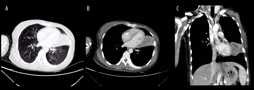
Figure 1
https://jours.isi-science.com/imageXml.php?i=amjcaserep-26-e947770-g001.jpg&idArt=947770&w=1000
Figure 1
Hình 1
Computed tomography scan showing solid lesions in the lingular segment of the left upper pulmonary lobe. Low-density cystic shadows were observed within the lesions, accompanied by mediastinal lymphadenopathy (A: lung window; B: mediastinal window; C: coronal reconstruction).
Phim chụp cắt lớp vi tính cho thấy các tổn thương dạng đặc ở phân thùy lưỡi của thùy trên phổi trái. Các bóng mờ dạng nang đậm độ thấp được quan sát thấy bên trong tổn thương, đi kèm với bệnh lý hạch trung thất (A: cửa sổ phổi; B: cửa sổ trung thất; C: tái tạo mặt phẳng đứng dọc [coronal]).
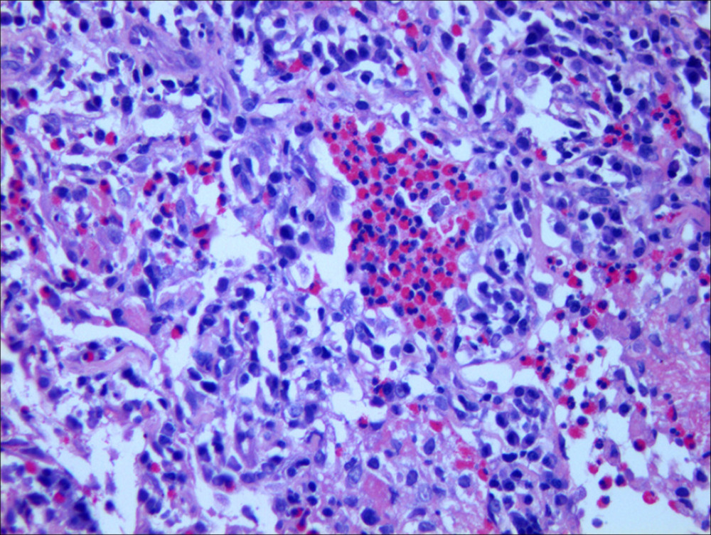
Figure 2
https://jours.isi-science.com/imageXml.php?i=amjcaserep-26-e947770-g002.jpg&idArt=947770&w=1000
Figure 2
Hình 2
Photomicrograph showing eosinophilic infiltration in the alveoli and interstitium, consistent with eosinophilic pneumonia. Images obtained after hematoxylin and eosin staining at 200× magnification.
Hình ảnh vi thể cho thấy sự thâm nhiễm bạch cầu ái toan trong các phế nang và mô kẽ, phù hợp với bệnh viêm phổi tăng bạch cầu ái toan. Hình ảnh thu được sau khi nhuộm hematoxylin và eosin ở độ phóng đại 200×.
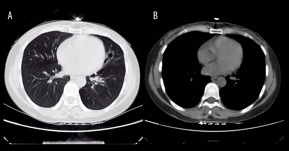
Figure 3
https://jours.isi-science.com/imageXml.php?i=amjcaserep-26-e947770-g003.jpg&idArt=947770&w=1000
Figure 3
Hình 3
Computed tomography scan demonstrating resolution of lesions in the lingular segment of the left upper lobe (A: lung window; B: mediastinal window).
Phim chụp cắt lớp vi tính cho thấy sự biến mất hoàn toàn của các tổn thương ở phân thùy lưỡi của thùy trên phổi trái (A: cửa sổ phổi; B: cửa sổ trung thất).
Có thể bạn quan tâm
-
Sự gia tăng thầm lặng: Một ca bệnh tăng canxi máu do tiêu cơ vân
The Silent Surge: A Case of Rhabdomyolysis-Induced Hypercalcemia
-
Hiệu quả của điều trị bằng thuốc so với giả dược hoặc không điều trị trong kiểm soát tình trạng mệt mỏi sau chấn thương sọ não (TBI)
Effects of pharmacological treatment compared with placebo or no treatment for fatigue management after traumatic brain injury (TBI)
-
Các can thiệp tâm lý xã hội nhằm hỗ trợ phụ nữ cai thuốc lá trong thai kỳ
Psychosocial interventions for supporting women to stop smoking in pregnancy
-
Khủng hoảng thai chết lưu tiềm ẩn ở châu Phi: Báo cáo mới vạch trần những lỗ hổng lớn về chính sách và dữ liệu
Africa’s hidden stillbirth crisis: new report exposes major policy and data gaps