Hội chứng chèn ép khoang thứ phát sau viêm tắc tĩnh mạch xanh đau ở bệnh nhân mắc bệnh Crohn

Compartment Syndrome Secondary to Phlegmasia Cerulea Dolens in a Patient With Crohn’s Disease

Saad Mohammed Alshahrani; Raghad Ibrahim Alkoblan; Reem Abdulaziz Alturki; Shahad Mohammed Alashgar. American Journal of Case Reports. doi: 10.12659/AJCR.948950. Open access.
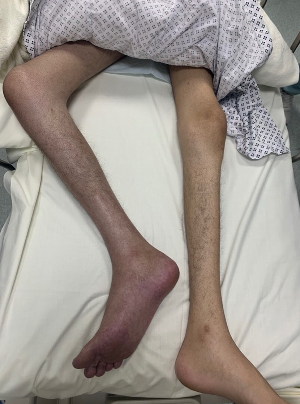
Right lower limb swelling and blue discoloration.
Figure 1
https://jours.isi-science.com/imageXml.php?i=amjcaserep-26-e948950-g001.jpg&idArt=948950&w=1000 Figure 1 Hình 1
Right lower limb swelling and blue discoloration.
Tình trạng sưng nề và đổi sang màu xanh ở chi dưới bên phải.
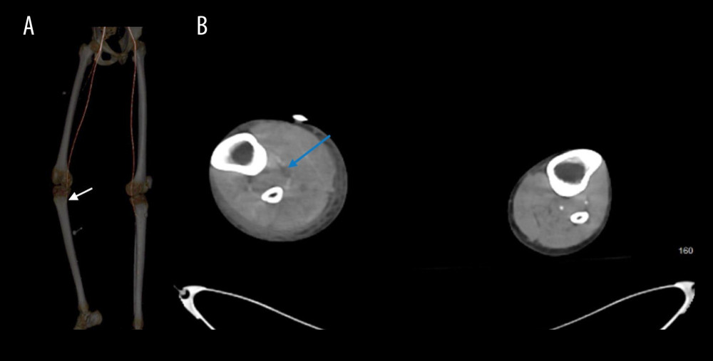
Computed tomography imaging demonstrating progressive non-opacification of the right infrageniculate arteries. (A) Lower limb computed tomography angiography revealing progressive non-opacification of the right infrageniculate arteries with distal loss of opacification (white arrow). (B) Abdominopelvic computed tomography (cross-sectional view) showing progressive non-opacification of the right infrageniculate arteries (blue arrow).
Figure 2
https://jours.isi-science.com/imageXml.php?i=amjcaserep-26-e948950-g002.jpg&idArt=948950&w=1000 Figure 2 Hình 2
Computed tomography imaging demonstrating progressive non-opacification of the right infrageniculate arteries. (A) Lower limb computed tomography angiography revealing progressive non-opacification of the right infrageniculate arteries with distal loss of opacification (white arrow). (B) Abdominopelvic computed tomography (cross-sectional view) showing progressive non-opacification of the right infrageniculate arteries (blue arrow).
Hình ảnh chụp cắt lớp vi tính cho thấy tình trạng không ngấm thuốc tiến triển của các động mạch dưới gối phải. (A) Chụp cắt lớp vi tính mạch máu chi dưới cho thấy tình trạng không ngấm thuốc tiến triển của các động mạch dưới gối phải kèm theo mất ngấm thuốc ở đoạn xa (mũi tên trắng). (B) Chụp cắt lớp vi tính bụng chậu (mặt cắt ngang) cho thấy tình trạng không ngấm thuốc tiến triển của các động mạch dưới gối phải (mũi tên xanh dương).
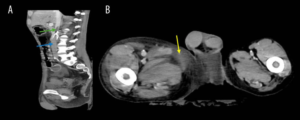
Abdominopelvic computed tomography imaging demonstrating venous thrombosis and associated soft tissue changes. (A) Abdominopelvic computed tomography (sagittal view) showing venous thrombosis extending above the inferior vena cava (IVC) filter (green arrow) into the infrahepatic segment (blue arrow). The IVC filter is visible immediately below the green arrow but not separately labeled. (B) Abdominopelvic computed tomography (cross-sectional view) showing asymmetrical soft tissue enlargement and congestion in the right thigh (yellow arrow).
Figure 3
https://jours.isi-science.com/imageXml.php?i=amjcaserep-26-e948950-g003.jpg&idArt=948950&w=1000 Figure 3 Hình 3
Abdominopelvic computed tomography imaging demonstrating venous thrombosis and associated soft tissue changes. (A) Abdominopelvic computed tomography (sagittal view) showing venous thrombosis extending above the inferior vena cava (IVC) filter (green arrow) into the infrahepatic segment (blue arrow). The IVC filter is visible immediately below the green arrow but not separately labeled. (B) Abdominopelvic computed tomography (cross-sectional view) showing asymmetrical soft tissue enlargement and congestion in the right thigh (yellow arrow).
Hình ảnh chụp cắt lớp vi tính bụng chậu cho thấy huyết khối tĩnh mạch và các thay đổi mô mềm kèm theo. (A) Chụp cắt lớp vi tính bụng chậu (mặt cắt đứng dọc) cho thấy huyết khối tĩnh mạch lan rộng lên trên lưới lọc tĩnh mạch chủ dưới (IVC) (mũi tên xanh lá) đi vào đoạn dưới gan (mũi tên xanh dương). Lưới lọc IVC có thể nhìn thấy ngay phía dưới mũi tên xanh lá nhưng không được dán nhãn riêng biệt. (B) Chụp cắt lớp vi tính bụng chậu (mặt cắt ngang) cho thấy tình trạng sưng phồng mô mềm không đối xứng và sung huyết ở đùi phải (mũi tên vàng).
Show
Hide
EN – VI
VI – EN

Abstract

Tóm tắt

BACKGROUND: Phlegmasia cerulea dolens is a rare, potentially fatal complication of deep vein thrombosis characterized by sudden limb swelling, pain, and cyanosis. The prevalence of venous thromboembolism is estimated to be 1.5 to 3.5 times higher in patients with inflammatory bowel disease than in the general population. Without prompt treatment, it may progress to venous gangrene, compartment syndrome, or circulatory shock. Crohn’s disease, a chronic inflammatory bowel disorder, increases the risk of thromboembolic events due to persistent inflammation and hypercoagulability, which can precipitate phlegmasia cerulea dolens.

BỐI CẢNH: Phlegmasia cerulea dolens (viêm tắc tĩnh mạch xanh đau) là một biến chứng hiếm gặp, có nguy cơ tử vong của bệnh huyết khối tĩnh mạch sâu, đặc trưng bởi tình trạng sưng chi, đau và tím tái khởi phát đột ngột. Tỷ lệ mắc thuyên tắc huyết khối tĩnh mạch ở bệnh nhân viêm ruột được ước tính cao hơn từ 1,5 đến 3,5 lần so với quần thể chung. Nếu không được điều trị kịp thời, bệnh có thể tiến triển thành hoại tử tĩnh mạch, hội chứng chèn ép khoang hoặc sốc tuần hoàn. Bệnh Crohn, một rối loạn viêm ruột mạn tính, làm tăng nguy cơ xảy ra các biến cố thuyên tắc huyết khối do tình trạng viêm kéo dài và tăng đông, điều này có thể đẩy nhanh sự tiến triển của phlegmasia cerulea dolens.

CASE REPORT: A 26-year-old male smoker with a history of fistulizing Crohn’s disease, nonadherence to his prescribed regimen, chronic pulmonary embolism, and prior inferior vena cava filter placement presented with acute right lower limb swelling and pain. Doppler ultrasonography revealed extensive iliofemoral deep vein thrombosis extending into the inferior vena cava; clinical evaluation confirmed compartment syndrome. Emergency fasciotomy and popliteal artery embolectomy restored circulation and prevented ischemic injury. Subsequently, the patient required a blood transfusion due to lower gastrointestinal bleeding, followed by an emergent exploratory laparotomy with right hemicolectomy, ileocecal resection, and ileostomy after colonoscopy failed to identify an active bleeding source. The patient recovered well postoperatively.

BÁO CÁO CA BỆNH: Một bệnh nhân nam 26 tuổi, có hút thuốc lá, với tiền sử bệnh Crohn thể rò, không tuân thủ phác đồ điều trị được kê đơn, thuyên tắc phổi mạn tính và đã từng đặt lưới lọc tĩnh mạch chủ dưới trước đó, nhập viện vì sưng và đau cấp tính ở chi dưới bên phải. Siêu âm Doppler cho thấy huyết khối tĩnh mạch sâu chậu-đùi lan rộng vào đến tĩnh mạch chủ dưới; đánh giá lâm sàng xác nhận có hội chứng chèn ép khoang. Phẫu thuật mở rạch giải áp khoang khẩn cấp và lấy huyết khối động mạch khoeo đã giúp khôi phục tuần hoàn và ngăn ngừa tổn thương thiếu máu cục bộ. Sau đó, bệnh nhân cần truyền máu do xuất huyết tiêu hóa dưới, tiếp theo là phẫu thuật mở bụng thăm dò khẩn cấp kết hợp cắt đại tràng phải, cắt đoạn hồi-manh tràng và làm hậu môn nhân tạo sau khi nội soi đại tràng không tìm thấy nguồn chảy máu hoạt động. Bệnh nhân hồi phục tốt sau phẫu thuật.

CONCLUSIONS

KẾT LUẬN

This case highlights the importance of early recognition and prompt management of phlegmasia cerulea dolens in patients with inflammatory bowel disease, particularly among younger individuals with an increased risk of thrombotic complications.

Ca bệnh này nhấn mạnh tầm quan trọng của việc nhận biết sớm và xử trí kịp thời tình trạng phlegmasia cerulea dolens ở bệnh nhân viêm ruột, đặc biệt là ở những người trẻ tuổi có nguy cơ cao gặp các biến chứng huyết khối.

Keywords: compartment syndromes, Crohn disease, fasciotomy, thrombosis

Từ khóa: hội chứng chèn ép khoang, bệnh Crohn, mở rạch giải áp khoang, huyết khối

Introduction

Đặt vấn đề

Phlegmasia cerulea dolens (PCD) is a rare, life-threatening manifestation of deep venous thrombosis that presents with sudden, extensive limb swelling, pain, and cyanosis, typically due to obstruction of the iliofemoral veins. Without timely treatment, PCD may progress to venous gangrene, compartment syndrome, or circulatory shock, highlighting the importance of early intervention 1. Risk factors include malignancy, hypercoagulable states, and venous stasis 2. PCD typically affects the left leg and is more common in older women 3. Due to its rarity, contemporary large-scale epidemiologic data are limited; most available evidence are derived from older case series and retrospective analyses. Nevertheless, PCD remains a high-risk condition: venous gangrene develops in approximately 40% to 60% of cases, and mortality rates range from 20% to 40% 4. These outcomes highlight the need for prompt recognition and aggressive management.

Phlegmasia cerulea dolens (PCD – viêm tắc tĩnh mạch xanh đau) là một biểu hiện hiếm gặp, đe dọa tính mạng của huyết khối tĩnh mạch sâu, biểu hiện bằng tình trạng sưng chi lan rộng, đau và tím tái khởi phát đột ngột, thường do tắc nghẽn các tĩnh mạch chậu-đùi. Nếu không được điều trị kịp thời, PCD có thể tiến triển thành hoại tử tĩnh mạch, hội chứng chèn ép khoang hoặc sốc tuần hoàn, điều này nhấn mạnh tầm quan trọng của việc can thiệp sớm 1. Các yếu tố nguy cơ bao gồm bệnh lý ác tính, tình trạng tăng đông và ứ trệ tĩnh mạch 2. PCD thường ảnh hưởng đến chân trái và phổ biến hơn ở phụ nữ lớn tuổi 3. Do tính chất hiếm gặp của bệnh, các dữ liệu dịch tễ học quy mô lớn hiện nay còn hạn chế; hầu hết các bằng chứng sẵn có đều bắt nguồn từ các chuỗi ca bệnh và phân tích hồi cứu cũ hơn. Tuy nhiên, PCD vẫn là một tình trạng nguy cơ cao: hoại tử tĩnh mạch phát triển ở khoảng 40% đến 60% số ca bệnh, và tỷ lệ tử vong dao động từ 20% to 40% 4. Những kết cục này làm nổi bật nhu cầu nhận biết nhanh chóng và xử trí tích cực.

Crohn’s disease, a form of chronic inflammatory bowel disease (IBD), is associated with an elevated risk of thromboembolic events due to chronic inflammation and hypercoagulability 5. The prevalence of venous thromboembolism (VTE) in patients with IBD is estimated to be 1.5 to 3.5 times higher than that in the general population 6. A long-term, population-based study in Manitoba, Canada showed that the overall incidence of VTE among individuals with IBD was 7.6%, compared with 3.3% among controls. Specifically, individuals with Crohn’s disease demonstrated an incidence of 8.4%, compared with 6.9% among individuals with ulcerative colitis 7. Similarly, in a prospective cohort study of 149 patients with IBD, 5% of those with Crohn’s disease had a documented history of thromboembolic events, including deep vein thrombosis (DVT), stroke, myocardial infarction, and pulmonary embolism (PE), further illustrating the clinical relevance of this risk 8. The mechanisms underlying the increased risk of VTE among individuals with IBD remain incompletely understood but appear multifactorial, involving dysregulation of the coagulation cascade, endothelial dysfunction, platelet activation, and impaired fibrinolysis. The chronic inflammatory state in Crohn’s disease alters both coagulation pathways and endothelial integrity, predisposing patients to events such as DVT and, in rare instances, PCD 6. Prompt diagnosis and intervention are essential to prevent irreversible ischemic damage in patients with PCD. Therapeutic options include anticoagulation, thrombolysis, and surgical thrombectomy; fasciotomy is sometimes required in cases complicated by compartment syndrome 9. This case report describes a rare instance of PCD in a young patient with Crohn’s disease that progressed to lower limb compartment syndrome and PE, emphasizing the importance of vigilance for thrombotic complications in this population.

Bệnh Crohn, một thể của bệnh viêm ruột mạn tính (IBD), có liên quan đến việc gia tăng nguy cơ xảy ra các biến cố thuyên tắc huyết khối do tình trạng viêm mạn tính và tăng đông 5. Tỷ lệ mắc thuyên tắc huyết khối tĩnh mạch (VTE) ở bệnh nhân IBD được ước tính cao hơn từ 1,5 đến 3,5 lần so với quần thể chung 6. Một nghiên cứu dựa trên quần thể dài hạn ở Manitoba, Canada cho thấy tỷ lệ bộc phát chung của VTE ở những cá nhân mắc IBD là 7,6%, so với 3,3% ở nhóm đối chứng. Cụ thể, những người mắc bệnh Crohn có tỷ lệ bộc phát là 8,4%, so với 6,9% ở những người bị viêm loét đại tràng 7. Tương tự, trong một nghiên cứu thuần tập tiến cứu trên 149 bệnh nhân IBD, 5% số bệnh nhân mắc bệnh Crohn có tiền sử được ghi nhận về các biến cố thuyên tắc huyết khối, bao gồm huyết khối tĩnh mạch sâu (DVT), đột quỵ, nhồi máu cơ tim và thuyên tắc phổi (PE), minh chứng rõ hơn cho mức độ liên quan lâm sàng của nguy cơ này 8. Cơ chế bệnh sinh đằng sau việc tăng nguy cơ VTE ở những người mắc IBD vẫn chưa được hiểu đầy đủ nhưng dường như do đa yếu tố, bao gồm sự mất điều hòa của chuỗi thác đông máu, rối loạn chức năng nội mạc, kích hoạt tiểu cầu và suy giảm ly giải fibrin. Tình trạng viêm mạn tính trong bệnh Crohn làm thay đổi cả con đường đông máu lẫn sự toàn vẹn của nội mạc, khiến bệnh nhân dễ gặp phải các biến cố như DVT và trong một số trường hợp hiếm gặp là PCD 6. Chẩn đoán và can thiệp kịp thời là điều thiết yếu để ngăn ngừa tổn thương thiếu máu cục bộ không thể đảo ngược ở bệnh nhân PCD. Các lựa chọn điều trị bao gồm dùng thuốc kháng đông, tiêu sợi huyết và phẫu thuật lấy huyết khối tĩnh mạch; phẫu thuật mở rạch giải áp khoang đôi khi được chỉ định trong các trường hợp biến chứng chèn ép khoang 9. Báo cáo ca bệnh này mô tả một trường hợp PCD hiếm gặp ở một bệnh nhân trẻ tuổi mắc bệnh Crohn tiến triển thành hội chứng chèn ép khoang chi dưới và PE, nhấn mạnh tầm quan trọng của việc cảnh giác với các biến chứng huyết khối ở quần thể bệnh nhân này.

Case Report

Báo cáo ca bệnh

A 26-year-old male smoker with a history of fistulizing Crohn’s disease diagnosed 8 years earlier presented with poor adherence to his prescribed medication regimen, which included azathioprine 50 mg orally once daily and infliximab 5 mg/kg intravenously each month. His medical history also included right pneumothorax secondary to right middle lobe cystic changes and empyema, which had been managed with a right middle lobectomy, as well as chronic recurrent PE for which an inferior vena cava (IVC) filter had been placed. The patient had initially been treated with apixaban when right segmental PE was diagnosed; after an instance of pneumothorax complicated by empyema and considering his ongoing nonadherence to anticoagulation, the decision was made to insert an IVC filter. During the present case, the patient attended the emergency department with a 1-day history of progressive swelling in the right lower limb, accompanied by pain and restricted range of motion. He reported decreased oral intake of solids and liquids, poor appetite, reduced activity, dizziness, and increased bloody diarrhea over the preceding 5 days.

Một bệnh nhân nam 26 tuổi, có hút thuốc lá, với tiền sử mắc bệnh Crohn thể rò được chẩn đoán trước đó 8 năm, nhập viện trong tình trạng tuân thủ kém đối với phác đồ thuốc được kê đơn, bao gồm azathioprine 50 mg uống một lần mỗi ngày và infliximab 5 mg/kg truyền tĩnh mạch mỗi tháng. Tiền sử y khoa của bệnh nhân cũng bao gồm tràn khí màng phổi phải thứ phát sau các thay đổi dạng kén ở thùy giữa phổi phải và tràn mủ màng phổi, vốn đã được xử trí bằng phẫu thuật cắt thùy giữa phổi phải, cũng như thuyên tắc phổi (PE) tái phát mạn tính đã được đặt lưới lọc tĩnh mạch chủ dưới (IVC). Ban đầu, bệnh nhân được điều trị bằng apixaban khi được chẩn đoán PE phân thùy phải; sau một đợt tràn khí màng phổi biến chứng tràn mủ màng phổi và xem xét tình trạng liên tục không tuân thủ điều trị kháng đông của bệnh nhân, quyết định đặt lưới lọc IVC đã được đưa ra. Trong ca bệnh này, bệnh nhân đến khoa cấp cứu với tiền sử sưng tiến triển ở chi dưới bên phải trong 1 ngày, kèm theo đau và giới hạn tầm vận động. Bệnh nhân báo cáo giảm lượng thức ăn đặc và lỏng nạp vào đường uống, chán ăn, giảm hoạt động, chóng mặt và tăng tiêu chảy ra máu trong 5 ngày trước đó.

On examination, the patient was hypotensive, with a blood pressure of 88/56 mmHg, and tachycardic, with a heart rate of 121 beats/min. Other vital signs were unremarkable, including a respiratory rate of 17 breaths/min, oxygen saturation of 100%, and temperature of 36.3°C. His body mass index was 14.5 kg/m2, with a height of 178 cm and weight of 46 kg. He received a 30 mL/kg bolus of intravenous normal saline, which improved his blood pressure. His remaining vital signs were stable. The right leg was diffusely swollen from the inguinal region to the foot, pale, and mottled with bluish discoloration (Figure 1$). Femoral pulses were palpable, and the right popliteal pulse was weakly palpable. In contrast, the posterior tibial and dorsalis pedis pulses were not palpable. The patient was unable to flex or extend his toes; he demonstrated no dorsiflexion or plantarflexion of the ankle against gravity. However, minimal movement was observed when gravity was eliminated, corresponding to a grade of 2/5 on the Medical Research Council Scale for Muscle Strength. Sensation in the affected limb was diminished.

Qua thăm khám, bệnh nhân bị hạ huyết áp, với huyết áp 88/56 mmHg, và nhịp tim nhanh, với tần số tim 121 lần/phút. Các dấu hiệu sinh tồn khác không có gì đặc biệt, bao gồm nhịp thở 17 lần/phút, độ bão hòa oxy 100% và nhiệt độ 36,3°C. Chỉ số khối cơ thể của bệnh nhân là 14,5 kg/m2, với chiều cao 178 cm và cân nặng 46 kg. Bệnh nhân được truyền một liều bolus tĩnh mạch dung dịch nước muối sinh lý 30 mL/kg, giúp cải thiện huyết áp. Các dấu hiệu sinh tồn còn lại của bệnh nhân ổn định. Chân phải bị sưng nề lan tỏa từ vùng bẹn đến bàn chân, nhợt nhạt và lốm đốm tím tái (Hình 1$). Mạch đùi có thể sờ thấy, và mạch khoeo phải sờ thấy yếu. Ngược lại, mạch chày sau và mạch mu chân không sờ thấy. Bệnh nhân không thể gập hoặc duỗi các ngón chân; không thể thực hiện động tác gập mu bàn chân hoặc gập lòng bàn chân của cổ chân chống lại trọng lực. Tuy nhiên, chuyển động tối thiểu đã được ghi nhận khi loại bỏ trọng lực, tương ứng với phân độ 2/5 trên Thang điểm Hội đồng Nghiên cứu Y khoa về Sức mạnh Cơ bắp. Cảm giác ở chi bị ảnh hưởng đã giảm đi.

Bedside and Doppler ultrasonography revealed substantial DVT in the iliofemoral vein. The Doppler study demonstrated triphasic waveforms in the femoral and popliteal arteries, monophasic waveforms in the posterior tibial artery, absence of detectable waves in the dorsalis pedis artery, and positive venous signals, indicating impaired arterial circulation and venous involvement. A continuous infusion of sodium heparin at 1000 U/mL was initiated, with subsequent prothrombin time and partial thromboplastin time values of 12.20 s and 34.20 s, respectively. Run-off computed tomography angiography revealed progressive non-opacification of the right infrageniculate arteries with distal loss of opacification; however, no distinct truncation or arterial embolus was identified in the right lower limb (Figure 2).

Siêu âm tại giường và siêu âm Doppler cho thấy huyết khối tĩnh mạch sâu (DVT) đáng kể ở tĩnh mạch chậu-đùi. Nghiên cứu Doppler cho thấy dạng sóng ba pha ở động mạch đùi và động mạch khoeo, dạng sóng một pha ở động mạch chày sau, không phát hiện được sóng ở động mạch mu chân và các tín hiệu tĩnh mạch dương tính, cho thấy sự suy giảm tuần hoàn động mạch và có sự tổn thương tĩnh mạch. Việc truyền liên tục heparin natri ở liều 1000 U/mL đã được bắt đầu, với các giá trị thời gian prothrombin và thời gian thromboplastin từng phần sau đó lần lượt là 12,20 giây và 34,20 giây. Chụp cắt lớp vi tính mạch máu (CTA) hạ lưu cho thấy tình trạng không ngấm thuốc tiến triển của các động mạch dưới gối phải kèm theo mất ngấm thuốc ở đoạn xa; tuy nhiên, không xác định được đoạn cắt cụ thể hoặc huyết khối động mạch nào ở chi dưới bên phải (Hình 2).

Pelvic computed tomography demonstrated extensive right lower limb venous thrombosis extending above the IVC filter into the infrahepatic IVC. The thrombosis was accompanied by asymmetrical soft tissue enlargement and congestion in the right thigh (Figure 3), prompting clinical evaluation for possible compartment syndrome. A diagnosis of acute right lower limb compartment syndrome secondary to extensive iliofemoral DVT and ischemia was established. Computed tomography venography to further assess thrombus extension was deferred to prevent delays in surgical management. Vascular surgery was consulted urgently. After computed tomography angiography confirmation of non-opacification of the right infrageniculate arteries, the patient underwent emergency fasciotomy using a double-incision technique and popliteal artery embolectomy. Blood flow was successfully restored; small thrombi were removed from the anterior and posterior tibial arteries. Leg color and tissue perfusion rapidly improved, and good biphasic Doppler signals were obtained. The patient tolerated the procedure well and was transferred to the intensive care unit for close observation. Shortly thereafter, he developed lower gastrointestinal bleeding. Emergent colonoscopy revealed no active bleeding site. Computed tomography angiography of the lower gastrointestinal tract demonstrated no evidence of active contrast extravasation or bleeding; thus, embolization was not performed. After appropriate resuscitation with blood transfusion, general surgery was consulted. The patient underwent emergent exploratory laparotomy with right hemicolectomy, ileocecal resection, and ileostomy. He remained in the intensive care unit for 7 days before transfer to the Gastroenterology ward. The patient was eventually discharged on low-molecular-weight heparin 40 mg twice daily and scheduled for IVC filter removal. He was ambulatory at discharge after a total hospital stay of 40 days.

Chụp cắt lớp vi tính vùng chậu cho thấy huyết khối tĩnh mạch chi dưới bên phải lan rộng, vượt lên trên lưới lọc IVC đi vào tĩnh mạch chủ dưới đoạn dưới gan. Huyết khối đi kèm với tình trạng sưng phồng mô mềm không đối xứng và sung huyết ở đùi phải (Hình 3), thôi thúc việc đánh giá lâm sàng về khả năng xảy ra hội chứng chèn ép khoang. Chẩn đoán hội chứng chèn ép khoang chi dưới bên phải cấp tính thứ phát sau DVT chậu-đùi lan rộng và thiếu máu cục bộ đã được thiết lập. Chụp cắt lớp vi tính tĩnh mạch (CTV) để đánh giá thêm sự lan rộng của huyết khối đã được hoãn lại nhằm tránh làm chậm trễ việc xử trí bằng phẫu thuật. Khoa phẫu thuật mạch máu đã được hội chẩn khẩn cấp. Sau khi kết quả CTA xác nhận tình trạng không ngấm thuốc của các động mạch dưới gối phải, bệnh nhân đã được phẫu thuật mở rạch giải áp khoang khẩn cấp bằng kỹ thuật hai đường rạch và phẫu thuật lấy huyết khối động mạch khoeo. Dòng máu đã được khôi phục thành công; các huyết khối nhỏ đã được loại bỏ khỏi động mạch chày trước và chày sau. Màu sắc da chân và sự tưới máu mô nhanh chóng được cải thiện, và thu được các tín hiệu Doppler hai pha tốt. Bệnh nhân dung nạp tốt với thủ thuật và được chuyển đến khoa hồi sức tích cực để theo dõi sát. Ngay sau đó, bệnh nhân xuất hiện tình trạng xuất huyết tiêu hóa dưới. Nội soi đại tràng khẩn cấp không phát hiện thấy vị trí chảy máu hoạt động nào. CTA đường tiêu hóa dưới cho thấy không có bằng chứng về việc thoát thuốc cản quang hoạt động hoặc chảy máu; do đó, thủ thuật nút mạch đã không được thực hiện. Sau khi hồi sức thích hợp bằng truyền máu, khoa ngoại tổng quát đã được hội chẩn. Bệnh nhân được phẫu thuật mở bụng thăm dò khẩn cấp kết hợp cắt đại tràng phải, cắt đoạn hồi-manh tràng và làm hậu môn nhân tạo. Bệnh nhân tiếp tục nằm tại khoa hồi sức tích cực trong 7 ngày trước khi chuyển sang khoa Tiêu hóa. Bệnh nhân cuối cùng được xuất viện với chỉ định dùng heparin trọng lượng phân tử thấp 40 mg hai lần mỗi ngày và được lên lịch hẹn để rút lưới lọc IVC. Bệnh nhân đã có thể tự đi lại lúc xuất viện sau tổng thời gian nằm viện là 40 ngày.

Discussion

Bàn luận

PCD is a rare but severe complication of DVT that may progress to compartment syndrome requiring urgent intervention 10. Compartment syndrome is usually diagnosed using a Stryker needle and comprises compartment pressure measurements of 30 mmHg or greater (normal pressures are typically below 10 mmHg); alternatively, it is regarded as a compartment pressure within 30 mmHg of the diastolic blood pressure 11. The pathophysiology of compartment syndrome in PCD involves venous hypertension secondary to obstruction of venous outflow, leading to interstitial edema and substantial fluid accumulation within the affected limb. This accumulation contributes to third-space fluid loss, which can precipitate hypovolemic shock. As compartment pressure rises, it may exceed arterial wall tension, causing arterial collapse. Subsequent elevation of interstitial and compartment pressures results in acute ischemia and venous gangrene, considerably increasing morbidity and mortality 12. Within 6 h, compartment pressures may rise by up to 16-fold due to severely impaired venous drainage 10. Although a few cases have been documented in the literature 10–14, compartment syndrome secondary to PCD remains rare. These reported cases matched the presentation of our patient, who exhibited classic features of PCD, including extensive swelling, cyanosis, and loss of motor and sensory function in the affected limb. The rapid progression of these symptoms, coupled with diminished distal pulses and imaging that confirmed extensive iliofemoral DVT with impaired arterial flow, was consistent with the expected pathophysiology. The urgent need for fasciotomy and embolectomy reflected the critical nature of compartment syndrome in this setting and highlighted the importance of early recognition and timely surgical intervention in improving patient outcomes in such rare cases.

PCD là một biến chứng hiếm gặp nhưng nghiêm trọng của DVT, có thể tiến triển thành hội chứng chèn ép khoang đòi hỏi phải can thiệp khẩn cấp 10. Hội chứng chèn ép khoang thường được chẩn đoán bằng kim Stryker và bao gồm các phép đo áp lực khoang từ 30 mmHg trở lên (áp lực bình thường thường là dưới 10 mmHg); cách khác, nó được xem là khi áp lực khoang nằm trong khoảng cách 30 mmHg so với huyết áp tâm trương 11. Cơ chế bệnh sinh của hội chứng chèn ép khoang trong PCD liên quan đến tình trạng tăng áp lực tĩnh mạch thứ phát do tắc nghẽn dòng chảy tĩnh mạch ra, dẫn đến phù kẽ và tích tụ một lượng dịch lớn trong chi bị ảnh hưởng. Sự tích tụ này góp phần gây mất dịch vào khoang thứ ba, có thể đẩy nhanh tình trạng sốc giảm thể tích. Khi áp lực khoang tăng lên, nó có thể vượt quá áp lực thành động mạch, gây xẹp động mạch. Sự gia tăng áp lực kẽ và áp lực khoang sau đó dẫn đến thiếu máu cục bộ cấp tính và hoại tử tĩnh mạch, làm tăng đáng kể tỷ lệ bệnh tật và tử vong 12. Trong vòng 6 giờ, áp lực khoang có thể tăng lên tới 16 lần do sự dẫn lưu tĩnh mạch bị suy giảm nghiêm trọng 10. Mặc dù một số ít ca bệnh đã được ghi nhận trong y văn 10–14, hội chứng chèn ép khoang thứ phát sau PCD vẫn là tình trạng hiếm gặp. Các ca bệnh được báo cáo này trùng khớp với bệnh cảnh của bệnh nhân chúng tôi, người đã thể hiện các đặc điểm kinh điển của PCD, bao gồm sưng nề lan rộng, tím tái và mất chức năng vận động cũng như cảm giác ở chi bị ảnh hưởng. Sự tiến triển nhanh chóng của các triệu chứng này, kết hợp với mạch ngoại vi giảm và hình ảnh học xác nhận DVT chậu-đùi lan rộng kèm theo suy giảm dòng chảy động mạch, là phù hợp với cơ chế bệnh sinh dự kiến. Nhu cầu khẩn cấp về phẫu thuật mở rạch giải áp khoang và lấy huyết khối đã phản ánh tính chất nghiêm trọng của hội chứng chèn ép khoang trong bối cảnh này, đồng thời nhấn mạnh tầm quan trọng của việc nhận biết sớm và can thiệp phẫu thuật kịp thời nhằm cải thiện kết cục của bệnh nhân trong những trường hợp hiếm gặp như vậy.

Intriguingly, younger patients with IBD have a greater likelihood of developing VTE relative to individuals without the condition. A nationwide Danish study demonstrated that both the incidence and relative risk of VTE unrelated to surgery, trauma, or pregnancy were higher among younger patients with IBD, especially those aged 20 years or younger, indicating a strong association between early-onset IBD and thrombotic risk 15. This finding aligns with the clinical history of our 26-year-old male patient with fistulizing Crohn’s disease, who experienced recurrent VTE episodes, including chronic PE, despite IVC filter placement. His poor adherence to immunosuppressive therapy and recent disease flare with bloody diarrhea likely further increased his thrombotic risk. Recurrent VTE in patients with IBD has also been shown to occur more frequently after cessation of anticoagulation, as demonstrated in the present case. A 2010 cohort study comparing patients with and without IBD who had a first unprovoked VTE episode revealed that the 5-year recurrence rate after discontinuing anticoagulation was significantly higher in the IBD group. Notably, younger age at the time of the initial thrombotic event was independently associated with an increased risk of recurrence, emphasizing the importance of early risk stratification and consideration of extended anticoagulation in this population 15. The recurrence of thrombosis after prior discontinuation of anticoagulation in our patient further supports findings in the literature, suggesting that prolonged or indefinite anticoagulation is warranted for young patients with IBD exhibiting high-risk profiles.

Điều thú vị là, những bệnh nhân trẻ tuổi mắc IBD có khả năng bộc phát VTE cao hơn so với những cá nhân không mắc tình trạng này. Một nghiên cứu trên toàn quốc ở Đan Mạch đã chứng minh rằng cả tỷ lệ bộc phát và nguy cơ tương đối của VTE không liên quan đến phẫu thuật, chấn thương hoặc thai kỳ đều cao hơn ở những bệnh nhân trẻ tuổi mắc IBD, đặc biệt là những người từ 20 tuổi trở xuống, cho thấy mối liên hệ mạnh mẽ giữa IBD khởi phát sớm và nguy cơ huyết khối 15. Phát hiện này phù hợp với tiền sử lâm sàng của bệnh nhân nam 26 tuổi mắc bệnh Crohn thể rò của chúng tôi, người đã trải qua các đợt VTE tái phát, bao gồm cả PE mạn tính, mặc dù đã được đặt lưới lọc IVC. Việc tuân thủ kém với liệu pháp ức chế miễn dịch và đợt bùng phát bệnh gần đây kèm theo tiêu chảy ra máu của bệnh nhân có khả năng đã làm tăng thêm nguy cơ huyết khối. VTE tái phát ở bệnh nhân IBD cũng được chứng minh là xảy ra thường xuyên hơn sau khi ngừng điều trị kháng đông, như đã được thể hiện trong ca bệnh này. Một nghiên cứu thuần tập năm 2010 so sánh các bệnh nhân có và không có IBD từng gặp biến cố VTE tự phát đầu tiên đã tiết lộ rằng tỷ lệ tái phát sau 5 năm sau khi ngừng thuốc kháng đông cao hơn đáng kể ở nhóm mắc IBD. Đáng chú ý, tuổi trẻ hơn tại thời điểm xảy ra biến cố huyết khối ban đầu có liên quan độc lập với việc tăng nguy cơ tái phát, nhấn mạnh tầm quan trọng của việc phân tầng nguy cơ sớm và xem xét kéo dài thời gian dùng thuốc kháng đông ở quần thể bệnh nhân này 15. Sự tái phát của huyết khối sau khi đã ngừng thuốc kháng đông trước đó ở bệnh nhân của chúng tôi càng củng cố thêm các phát hiện trong y văn, gợi ý rằng việc điều trị kháng đông kéo dài hoặc vô thời hạn là có cơ sở đối với những bệnh nhân trẻ tuổi mắc IBD có hồ sơ nguy cơ cao.

Patients with PCD usually present with blue or purple discoloration of the affected limb, accompanied by swelling, pain, and, in severe cases, loss of pulses 16. Because of the anatomical relationship in which the right iliac artery crosses over the left iliac vein, most cases involve the lower extremities; the left leg is more frequently affected than the right 12. However, our patient presented with right lower limb ischemia secondary to extensive iliofemoral DVT. Gong et al evaluated the association between the location of lower extremity DVT and PE. They found that right-sided DVT was a more frequent cause of PE than left-sided DVT, consistent with our patient’s history of chronic PE 17. Endothelial wall damage can occur through various mechanisms. Direct causes include mechanical disruption during procedures such as catheter insertion 18. Notably, our patient had undergone IVC filter placement, which was intended to prevent recurrent PE due to his history of chronic embolic disease. As highlighted in prior studies, the present case underscores the delicate balance between the preventive role of IVC filters and their potential to increase VTE risk, making close monitoring essential 19. Indirect endothelial wall damage may occur through inflammatory mechanisms 18. In our case, the patient’s Crohn’s disease, a form of IBD, further increased his risk of thrombosis. IBD enhances the likelihood of VTE by approximately 1.5 to 3.5 times and is associated with considerable morbidity and mortality 20,21. Endothelial dysfunction is a central feature of IBD pathogenesis, characterized by an imbalance between vasodilatory and vasoconstrictive mediators, typically driven by inflammation 22. This imbalance alters the structural and functional properties of endothelial cells 23. The resulting prothrombotic state in IBD increases susceptibility to severe thrombotic complications such as PCD, as demonstrated in the present case 24. Our patient demonstrated malnutrition, as reflected by his low body mass index. In severe IBD, particularly ulcerative colitis, malnutrition and nutrient deficiencies can impair coagulation factor synthesis, thereby predisposing patients to VTE 25.

Bệnh nhân mắc PCD thường nhập viện với tình trạng da đổi sang màu xanh hoặc tím ở chi bị ảnh hưởng, đi kèm với sưng, đau và trong các trường hợp nghiêm trọng là mất mạch 16. Do mối tương quan về mặt giải phẫu khi động mạch chậu phải bắt chéo phía trên tĩnh mạch chậu trái, hầu hết các ca bệnh đều liên quan đến các chi dưới; chân trái thường bị ảnh hưởng thường xuyên hơn chân phải 12. Tuy nhiên, bệnh nhân của chúng tôi lại nhập viện với tình trạng thiếu máu cục bộ chi dưới bên phải thứ phát sau DVT chậu-đùi lan rộng. Gong và các cộng sự đã đánh giá mối liên hệ giữa vị trí của DVT chi dưới và PE. Họ tìm thấy rằng DVT bên phải là nguyên nhân gây ra PE thường gặp hơn so với DVT bên trái, phù hợp với tiền sử PE mạn tính của bệnh nhân chúng tôi 17. Tổn thương thành nội mạc có thể xảy ra thông qua nhiều cơ chế khác nhau. Các nguyên nhân trực tiếp bao gồm sự phá vỡ cơ học trong các thủ thuật như đặt ống thông 18. Đáng chú ý là bệnh nhân của chúng tôi đã được đặt lưới lọc IVC, vốn nhằm mục đích ngăn ngừa PE tái phát do tiền sử bệnh thuyên tắc mạn tính của mình. Như đã được nhấn mạnh trong các nghiên cứu trước đây, ca bệnh hiện tại làm nổi bật sự cân bằng mong manh giữa vai trò phòng ngừa của lưới lọc IVC và tiềm năng làm tăng nguy cơ VTE của chúng, khiến việc theo dõi sát sao trở nên thiết yếu 19. Tổn thương thành nội mạc gián tiếp có thể xảy ra thông qua các cơ chế viêm 18. Trong trường hợp của chúng tôi, bệnh Crohn của bệnh nhân, một thể của IBD, đã làm tăng thêm nguy cơ huyết khối của anh ấy. IBD làm tăng khả năng xảy ra VTE khoảng 1,5 đến 3,5 lần và có liên quan đến tỷ lệ bệnh tật cũng như tử vong đáng kể 20,21. Rối loạn chức năng nội mạc là một đặc điểm trung tâm trong cơ chế bệnh sinh của IBD, được đặc trưng bởi sự mất cân bằng giữa các chất trung gian giãn mạch và co mạch, thường do tình trạng viêm thúc đẩy 22. Sự mất cân bằng này làm thay đổi các đặc tính cấu trúc và chức năng của tế bào nội mạc 23. Tình trạng tăng đông hệ quả trong IBD làm tăng tính nhạy cảm đối với các biến chứng huyết khối nghiêm trọng như PCD, như đã được chứng minh trong ca bệnh này 24. Bệnh nhân của chúng tôi cho thấy tình trạng suy dinh dưỡng, phản ánh qua chỉ số khối cơ thể thấp. Trong IBD nghiêm trọng, đặc biệt là viêm loét đại tràng, tình trạng suy dinh dưỡng và thiếu hụt chất dinh dưỡng có thể làm giảm tổng hợp các yếu tố đông máu, từ đó khiến bệnh nhân dễ bị VTE 25.

Endovascular therapies such as percutaneous mechanical thrombectomy and catheter-directed thrombolysis, as well as surgical intervention, may be considered treatment options. However, these approaches carry risks, including periprocedural PE and venous endothelial injury 26. The severity of ischemia determines whether an endovascular or surgical approach is most appropriate 27. In the present case, the patient underwent emergency fasciotomy combined with popliteal artery embolectomy. The decision to perform fasciotomy was driven by the need for rapid decompression of elevated compartment pressure, which, if not promptly managed, can result in irreversible ischemic damage to the affected tissues 28. Embolectomy addressed the arterial occlusion via direct removal of thrombi from the popliteal artery, restoring blood flow by means of an invasive but definitive procedure.

Các liệu pháp nội mạch như lấy huyết khối bằng cơ học qua da và tiêu sợi huyết qua ống thông, cũng như can thiệp phẫu thuật, có thể được xem xét làm các lựa chọn điều trị. Tuy nhiên, các phương pháp tiếp cận này mang lại những rủi ro, bao gồm PE trong quá trình làm thủ thuật và tổn thương nội mạc tĩnh mạch 26. Mức độ nghiêm trọng của tình trạng thiếu máu cục bộ sẽ quyết định liệu phương pháp tiếp cận nội mạch hay phẫu thuật là phù hợp nhất 27. Trong ca bệnh này, bệnh nhân đã trải qua phẫu thuật mở rạch giải áp khoang khẩn cấp kết hợp với lấy huyết khối động mạch khoeo. Quyết định thực hiện mở rạch giải áp khoang được thúc đẩy bởi nhu cầu giảm áp nhanh chóng áp lực khoang đang tăng cao, điều mà nếu không được xử trí kịp thời có thể dẫn đến tổn thương thiếu máu cục bộ không thể đảo ngược đối với các mô bị ảnh hưởng 28. Phẫu thuật lấy huyết khối đã giải quyết tình trạng tắc nghẽn động mạch thông qua việc loại bỏ trực tiếp các cục máu đông khỏi động mạch khoeo, khôi phục dòng máu bằng một thủ thuật xâm lấn nhưng mang tính triệt để.

Conclusions

Kết luận

PCD with compartment syndrome is rare but associated with high morbidity and mortality among patients with IBD, particularly younger individuals who exhibit an increased risk of VTE due to chronic inflammation and coagulation abnormalities. This risk is driven by chronic systemic inflammation, dysregulation of the coagulation cascade, endothelial dysfunction, platelet activation, and impaired fibrinolysis. Right-sided DVT, as demonstrated in the present case, may further enhance the risk of PE. Prompt recognition and aggressive management are essential to restore venous flow, prevent limb loss, and address the added complication of compartment syndrome, which requires urgent decompression and close monitoring.

PCD (viêm tắc tĩnh mạch xanh đau) kèm theo hội chứng chèn ép khoang là tình trạng hiếm gặp nhưng có liên quan đến tỷ lệ bệnh tật và tử vong cao ở các bệnh nhân mắc IBD, đặc biệt là những người trẻ tuổi – đối tượng vốn có nguy cơ bộc phát VTE cao hơn do tình trạng viêm mạn tính và các bất thường về đông máu. Nguy cơ này được thúc đẩy bởi tình trạng viêm hệ thống mạn tính, sự mất điều hòa của chuỗi thác đông máu, rối loạn chức năng nội mạc, kích hoạt tiểu cầu và suy giảm ly giải fibrin. DVT bên phải, như đã được chứng minh trong ca bệnh này, có thể làm tăng thêm nguy cơ xảy ra PE. Việc nhận biết nhanh chóng và xử trí tích cực là điều thiết yếu để khôi phục dòng chảy tĩnh mạch, ngăn ngừa mất chi, đồng thời giải quyết biến chứng đi kèm của hội chứng chèn ép khoang – tình trạng đòi hỏi phải giải áp khẩn cấp và theo dõi sát sao.

Reference

  1. Ogeng’o JA, Ongera D, Sinkeet SR, Phlegmasia cerulea dolens: A review of pathophysiology and management strategies: Int J Vasc Med, 2021; 2021; 7809435
  2. Hussain MA, Aljabri B, Al-Omran M, The importance of recognizing phlegmasia cerulea dolens in patients with high-risk conditions: J Vasc Surg Venous Lymphat Disord, 2018; 6(1); 32-39
  3. Chinsakchai K, Ten Duis K, Moll FL, Phlegmasia cerulea dolens: Incidence and outcomes in older adults: Eur J Vasc Endovasc Surg, 2020; 59(2); 335-42
  4. Klok FA, Huisman MV, Seeking optimal treatment for phlegmasia cerulea dolens: Thromb Res, 2013; 131(4); 372-73
  5. Koutroubakis IE, Tsiolakidou G, Koumoutsos I, Risk of thrombosis in inflammatory bowel disease: A comprehensive review: World J Gastroenterol, 2015; 21(3); 793-802
  6. Cheng K, Faye AS, Venous thromboembolism in inflammatory bowel disease: World J Gastroenterol, 2020; 26(12); 1231-41
  7. Bernstein CN, Nugent Z, Singh H, Persistently high rate of venous thromboembolic disease in inflammatory bowel disease: A population-based study: Am J Gastroenterol, 2021; 116(7); 1476-84
  8. Carvalho AC, Pinho J, Cancela E, Inflammatory bowel disease and thromboembolic events: A clot to learn: Ther Adv Gastroenterol, 2022; 15; 17562848221100626
  9. Hozumi J, Hozumi T, Takahashi Y, Emergency management of phlegmasia cerulea dolens with compartment syndrome: A case report: Am J Emerg Med, 2019; 37(12); 2276e5-e8
  10. Aydemir B, Hoyle C, Hakmeh W, Phlegmasia cerulea dolens causing compartment syndrome: Am J Emerg Med, 2022; 61; 234e1-e3
  11. Davidson AL, Sutherland MA, Siska RC, Janis JE, Practical review on the contemporary diagnosis and management of compartment syndrome: Plast Reconstr Surg Glob Open, 2024; 12(3); e5637
  12. Gardella L, Faulk JB, Phlegmasia alba and cerulea dolens [Updated 2022 Oct 3]: StatPearls [Internet], 2025, Treasure Island (FL), StatPearls Publishing Available from: https://www.ncbi.nlm.nih.gov/books/NBK563137/
  13. Chaochankit W, Akaraborworn O, Phlegmasia cerulea dolens with compartment syndrome: Ann Vasc Dis, 2018; 11(3); 355-57
  14. Dah K, Ogbu IR, Ngwudike C, Tripathi M, Right lower extremity phlegmasia cerulea dolens due to iliotibial vein thrombosis with compartment syndrome and circulatory shock: Case report and review: Cureus, 2022; 14(12); e32364
  15. Coremans L, Strubbe B, Peeters H, Venous thromboembolism in patients with inflammatory bowel disease: Review of literature and practical algorithms: Acta Gastroenterol Belg, 2021; 84(1); 79-85
  16. Zhang Q, Yang L, Chang L, Analysis and literature review of deep vein thrombosis related phlegmasia cerulea dolens diagnosis in critically patients: Case Rep Clin Med, 2023; 12(8); 312-23
  17. Gong S, Lee EJ, Kim JS, Association between laterality and location of deep vein thrombosis of lower extremity and pulmonary embolism: Vasc Specialist Int, 2021; 37; 12
  18. Ashorobi D, Ameer MA, Fernandez R, Thrombosis [Updated 2024 Feb 12]: StatPearls [Internet], 2025, Treasure Island (FL), StatPearls Publishing Available from: https://www.ncbi.nlm.nih.gov/books/NBK538430/
  19. Grewal S, Chamarthy MR, Kalva SP, Complications of inferior vena cava filters: Cardiovasc Diagn Ther, 2016; 6(6); 632-41
  20. Chung WS, Lin CL, Hsu WH, Kao CH, Inflammatory bowel disease increases the risks of deep vein thrombosis and pulmonary embolism in the hospitalized patients: A nationwide cohort study: Thromb Res, 2015; 135(3); 492-96
  21. Kohoutova D, Moravkova P, Kruzliak P, Bures J, Thromboembolic complications in inflammatory bowel disease: J Thromb Thrombolysis, 2015; 39(4); 489-98
  22. Cibor D, Domagala-Rodacka R, Rodacki T, Endothelial dysfunction in inflammatory bowel diseases: Pathogenesis, assessment and implications: World J Gastroenterol, 2016; 22(3); 1067-77
  23. Boussi L, Safaya A, Goyal A, “Inflammatory bowel disease – not just the bowel’s bane”: Peripheral arterial and venous thrombosis in a patient with Crohn disease: Vasc Endovascular Surg”, 2020; 54(7); 646-49
  24. Zezos P, Kouklakis G, Saibil F, Inflammatory bowel disease and thromboembolism: World J Gastroenterol, 2014; 20(38); 13863-78
  25. Cryer B, The risks of deep vein thrombosis in patients with ulcerative colitis and COVID-19: Proc (Bayl Univ Med Cent), 2024; 37(2); 248-49
  26. Šimon S, Roman S, Case report of simultaneous phlegmasia cerulea dolens and acute limb ischemia: Int J Surg Case Rep, 2024; 125; 110596
  27. Kou CJ, Batzlaff C, Bezzant ML, Sjulin T, Phlegmasia cerulea dolens: A life-threatening manifestation of deep vein thrombosis: Cureus, 2020; 12(6); e8587
  28. Torlincasi AM, Lopez RA, Waseem M, Acute compartment syndrome [Updated 2023 Jan 16]: StatPearls [Internet], 2025, Treasure Island (FL), StatPearls Publishing Available from:https://www.ncbi.nlm.nih.gov/books/NBK448124/

Có thể bạn quan tâm