Ca lâm sàng
Phát hiện dị dạng động tĩnh mạch phế quản trước khi triệt đốt bằng catheter đối với cuồng nhĩ và rung nhĩ kịch phát: Báo cáo ca lâm sàng
Bronchial Arteriovenous Malformation Detection Prior to Catheter Ablation for Paroxysmal Atrial Flutter and Fibrillation: A Case Report
Alexandra Arvanitaki; Marios Georgios Bantidos; Theodora Zaglavara; Lydia Kalogeropoulou; Athanasios Samaras; Athina Nasoufidou; Efstratios Karagiannidis; Georgios Stavropoulos; Apostolos Tzikas; Nikolaos Fragakis. American Journal of Case Reports. doi: 10.12659/AJCR.949879. Open access.
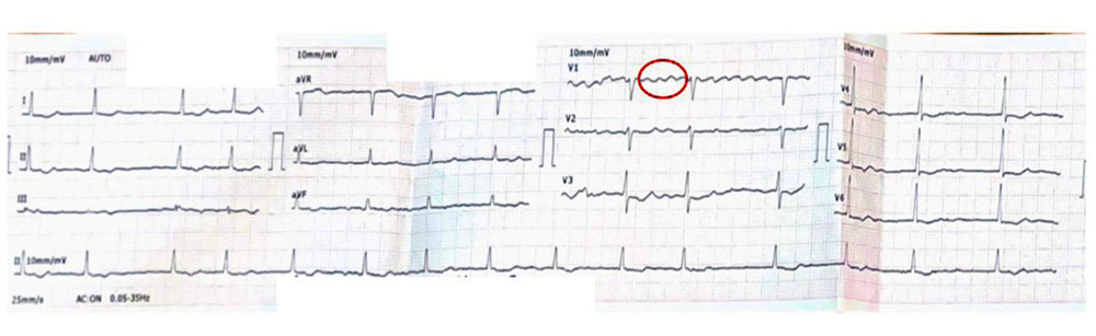
Figure 1
https://jours.isi-science.com/imageXml.php?i=amjcaserep-26-e949879-g001.jpg&idArt=949879&w=1000
Figure 1
Hình 1
Electrocardiogram of paroxysmal anticlockwise re-entry atrial flutter, indicating right isthmus dependenceA “sawtooth” pattern of inverted flutter waves is present in leads II, III; an augmented vector foot and upright flutter waves are present in lead V1 (red circle).
Điện tâm đồ của cơn cuồng nhĩ kịch phát do vòng vào lại ngược chiều kim đồng hồ, cho thấy sự phụ thuộc vào eo phải. Dạng hình “răng cưa” của các sóng cuồng nhĩ đảo ngược hiện diện ở các chuyển đạo II, III; một chuyển đạo aVF và các sóng cuồng nhĩ thẳng đứng hiện diện ở chuyển đạo V1 (vòng tròn đỏ).
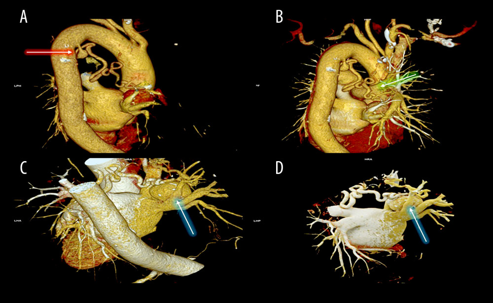
Figure 2
https://jours.isi-science.com/imageXml.php?i=amjcaserep-26-e949879-g002.jpg&idArt=949879&w=1000
Figure 2
Hình 2
Three-dimensional computed tomography volume-rendering technique reconstruction of the bronchial arteriovenous malformation(A) Meandering course of the bronchial artery with a small aneurysm indicated near its origin from the thoracic aorta (red arrow). (B) The bronchial artery drains into the right pulmonary artery through a small branch (green arrow). (C, D) A larger saccular aneurysm with mural calcifications is noted distally along the bronchial artery near the left atrium (blue arrows).
Tái tạo hình ảnh dị dạng động tĩnh mạch phế quản bằng kỹ thuật dựng hình thể tích từ chụp cắt lớp vi tính 3 chiều. (A) Đường đi ngoằn ngoèo của động mạch phế quản với một túi phình nhỏ được chỉ ra ở gần vị trí xuất phát từ động mạch chủ ngực (mũi tên đỏ). (B) Động mạch phế quản đổ vào động mạch phổi phải thông qua một nhánh nhỏ (mũi tên xanh lá). (C, D) Một túi phình hình túi lớn hơn với các tổn thương vôi hóa thành mạch được ghi nhận ở đoạn xa dọc theo động mạch phế quản, gần tâm nhĩ trái (mũi tên xanh dương).
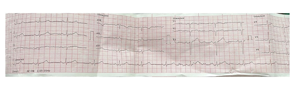
Figure 3
https://jours.isi-science.com/imageXml.php?i=amjcaserep-26-e949879-g003.jpg&idArt=949879&w=1000
Figure 3
Hình 3
Resting electrocardiogram in sinus rhythm (55 beats/min)No cardiac pathology is present.
Điện tâm đồ lúc nghỉ ở nhịp xoang (55 lần/phút). Không có bệnh lý tim mạch.
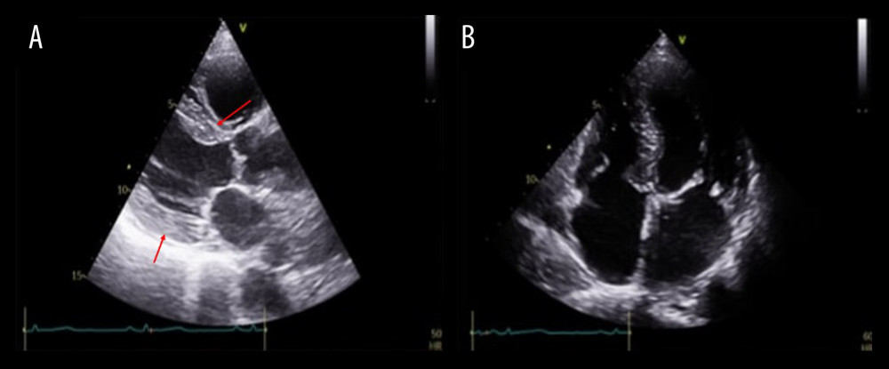
Figure 4
https://jours.isi-science.com/imageXml.php?i=amjcaserep-26-e949879-g004.jpg&idArt=949879&w=1000
Figure 4
Hình 4
Transthoracic echocardiography(A) Parasternal long-axis view shows mild left ventricular hypertrophy (red arrows indicate interventricular septum and posterior wall of the left ventricle) due to arterial hypertension, with normal left ventricular size. (B) Apical 4-chamber view depicts normal biatrial size. Left ventricular function is within normal range.
Siêu âm tim qua thành ngực. (A) Mặt cắt trục dọc cạnh ức cho thấy phì đại tâm thất trái nhẹ (mũi tên đỏ chỉ vách liên thất và thành sau của tâm thất trái) do tăng huyết áp động mạch, với kích thước tâm thất trái bình thường. (B) Mặt cắt 4 buồng từ mỏm mô tả kích thước hai tâm nhĩ bình thường. Chức năng tâm thất trái nằm trong giới hạn bình thường.
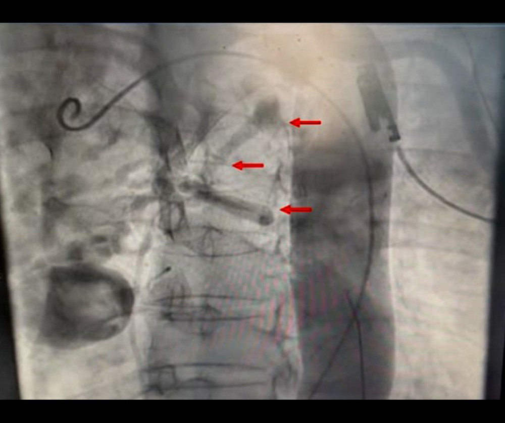
Figure 5
https://jours.isi-science.com/imageXml.php?i=amjcaserep-26-e949879-g005.jpg&idArt=949879&w=1000
Figure 5
Hình 5
Invasive aortography assessment of the bronchial arteriovenous malformation (BAVM)Selective aortography demonstrates the BAVM arising from the descending thoracic aorta (red arrows). Opacification of drainage into the pulmonary artery is limited, likely due to contrast dilution within the pulmonary circulation.
Đánh giá dị dạng động tĩnh mạch phế quản (BAVM) bằng chụp động mạch chủ xâm lấn. Chụp động mạch chủ chọn lọc cho thấy BAVM xuất phát từ động mạch chủ ngực xuống (mũi tên đỏ). Sự hiện hình của dòng đổ vào động mạch phổi bị hạn chế, có thể là do thuốc cản quang bị pha loãng trong tuần hoàn phổi.
Có thể bạn quan tâm
-
Sarcoidosis thần kinh và sarcoidosis tim đồng mắc: Một lộ trình chẩn đoán đặc biệt
Concomitant Neurosarcoidosis and Cardiac Sarcoidosis: A Peculiar Path to Diagnosis
-
Hiệu quả của các can thiệp kích thích hoặc phản hồi sinh học trong kiểm soát tình trạng mệt mỏi sau chấn thương sọ não (TBI)
Effects of stimulation or biofeedback interventions for fatigue management after traumatic brain injury (TBI)
-
Thử nghiệm ngẫu nhiên về việc sử dụng Prednisolone bổ trợ trong điều trị bệnh Kawasaki
Randomized Trial of Adjunctive Prednisolone for Kawasaki Disease
-
Sốt thung lũng nứt: nó là gì, cách nó lây lan và cách ngăn chặn nó
Rift Valley fever: what it is, how it spreads and how to stop it