Ca lâm sàng
Đau cổ đơn độc là một biểu hiện không điển hình của bóc tách động mạch chủ cấp tính type A: Báo cáo ca lâm sàng
Isolated Neck Pain as an Atypical Manifestation of Type A Acute Aortic Dissection: A Case Report
Łukasz Turek; Jacek Kurzawski; Łukasz Zandecki; Małgorzata Zachura; Agata Kundera-Mądro; Marianna Janion. American Journal of Case Reports. doi: 10.12659/AJCR.949758. Open access.
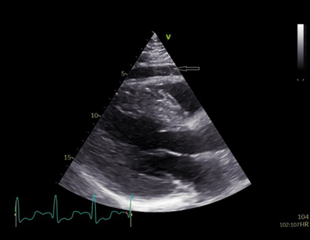
Figure 1
https://jours.isi-science.com/imageXml.php?i=amjcaserep-26-e949758-g001.jpg&idArt=949758&w=1000
Figure 1
Hình 1
Two-dimensional transthoracic echocardiography. The parasternal long-axis view demonstrates left ventricular hypertrophy and regional pericardial effusion (arrow) anterior to the right ventricle.
Siêu âm tim qua thành ngực hai chiều (2D). Mặt cắt dọc cạnh ức minh họa tình trạng phì đại thất trái và tràn dịch màng tim khu trú (mũi tên) ở phía trước thất phải.
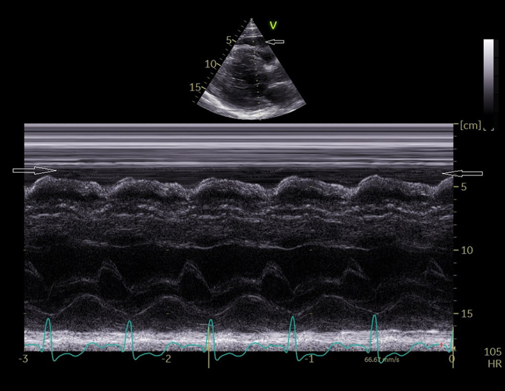
Figure 2
https://jours.isi-science.com/imageXml.php?i=amjcaserep-26-e949758-g002.jpg&idArt=949758&w=1000
Figure 2
Hình 2
M-mode transthoracic echocardiography. The parasternal long-axis view shows regional pericardial effusion (arrows) anterior to the right ventricle.
Siêu âm tim qua thành ngực kiểu M (M-mode). Mặt cắt dọc cạnh ức hiển thị tình trạng tràn dịch màng tim khu trú (các mũi tên) ở phía trước thất phải.
![Two-dimensional transthoracic echocardiography. The suprasternal view of the aorta reveals 2 vascular lumina (true [arrow] and false [arrowhead]) separated by an intimal flap, indicative of ascending aortic dissection extending into the proximal aortic arch.](https://jours.isi-science.com/imageXml.php?i=amjcaserep-26-e949758-g003.jpg&idArt=949758&w=1000)
Figure 3
https://jours.isi-science.com/imageXml.php?i=amjcaserep-26-e949758-g003.jpg&idArt=949758&w=1000
Figure 3
Hình 3
Two-dimensional transthoracic echocardiography. The suprasternal view of the aorta reveals 2 vascular lumina (true [arrow] and false [arrowhead]) separated by an intimal flap, indicative of ascending aortic dissection extending into the proximal aortic arch.
Siêu âm tim qua thành ngực hai chiều (2D). Mặt cắt trên hõm ức của động mạch chủ phát hiện 2 lòng mạch (lòng thật [mũi tên] và lòng giả [đầu mũi tên]) được phân tách bởi một vạt nội mạc, biểu hiện của tình trạng bóc tách động mạch chủ lên lan vào phần gần của quai động mạch chủ.
![Two-dimensional transthoracic echocardiography. The suprasternal view of the aorta shows 2 vascular lumina (true [arrow] and false [arrowhead]) separated by an intimal flap, indicating an ascending aortic dissection extending into the proximal aortic arch.](https://jours.isi-science.com/imageXml.php?i=amjcaserep-26-e949758-g004.jpg&idArt=949758&w=1000)
Figure 4
https://jours.isi-science.com/imageXml.php?i=amjcaserep-26-e949758-g004.jpg&idArt=949758&w=1000
Figure 4
Hình 4
Two-dimensional transthoracic echocardiography. The suprasternal view of the aorta shows 2 vascular lumina (true [arrow] and false [arrowhead]) separated by an intimal flap, indicating an ascending aortic dissection extending into the proximal aortic arch.
Siêu âm tim qua thành ngực hai chiều (2D). Mặt cắt trên hõm ức của động mạch chủ cho thấy 2 lòng mạch (lòng thật [mũi tên] và lòng giả [đầu mũi tên]) được phân tách bởi một vạt nội mạc, biểu thị một trường hợp bóc tách động mạch chủ lên lan vào phần gần của quai động mạch chủ.
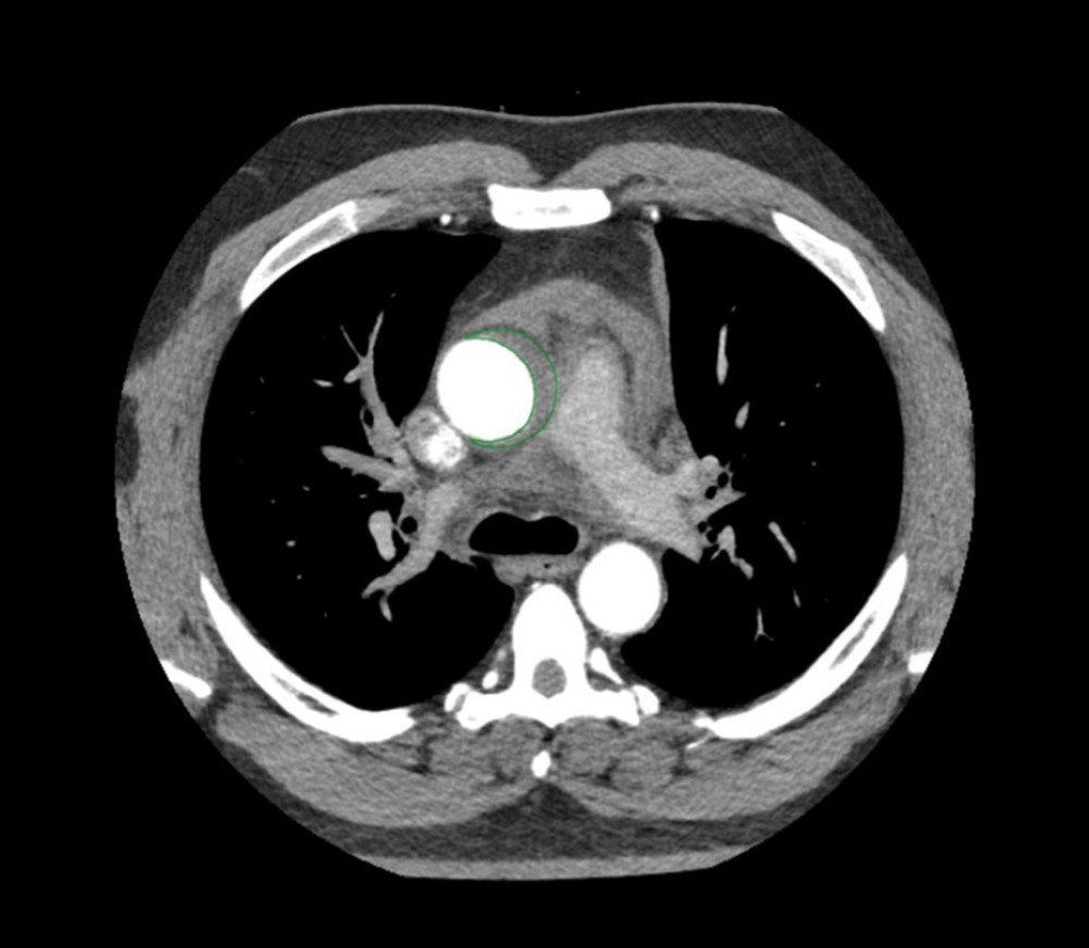
Figure 5
https://jours.isi-science.com/imageXml.php?i=amjcaserep-26-e949758-g005.jpg&idArt=949758&w=1000
Figure 5
Hình 5
Contrast-enhanced computed tomography with axial reconstruction suggestive of Stanford Type A acute aortic dissection (gray region inside the green line).
Chụp cắt lớp vi tính (CT) có cản quang với hình ảnh tái tạo theo mặt phẳng ngang (axial) gợi ý tình trạng bóc tách động mạch chủ cấp tính Stanford Type A (vùng màu xám bên trong đường màu xanh lá cây).
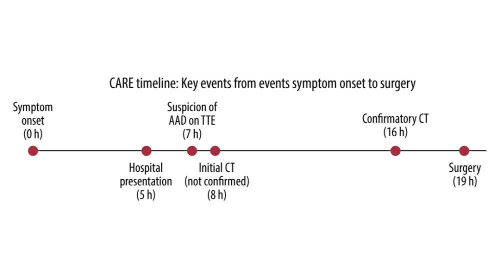
Figure 6
https://jours.isi-science.com/imageXml.php?i=amjcaserep-26-e949758-g006.jpg&idArt=949758&w=1000
Figure 6
Hình 6
CARE timeline illustrating key diagnostic and therapeutic events from symptom onset to surgery. CARE – CAse REport guidelines; AAD – acute aortic dissection; TTE – transthoracic echocardiography; CT – computed tomography.
Dòng thời gian CARE minh họa các sự kiện chẩn đoán và điều trị chính từ khi khởi phát triệu chứng đến khi phẫu thuật. CARE – Hướng dẫn Báo cáo Ca lâm sàng; AAD – bóc tách động mạch chủ cấp tính; TTE – siêu âm tim qua thành ngực; CT – chụp cắt lớp vi tính.
Có thể bạn quan tâm
-
Tự sát bằng nhiều phát súng vào ngực trong một vụ án giết người – tự tử: Một trường hợp bất thường
Thoracic Multiple-Gunshot Suicide in a Homicide–Suicide Incident: An Unusual Case
-
Phẫu thuật nội soi so với phẫu thuật mở trong điều trị thoát vị bẹn ở trẻ em
Laparoscopic versus open repair for pediatric inguinal hernia
-
Các can thiệp tâm lý xã hội nhằm hỗ trợ phụ nữ cai thuốc lá trong thai kỳ
Psychosocial interventions for supporting women to stop smoking in pregnancy
-
Tòa án Tối cao duy trì quyền tiếp cận mifepristone qua dịch vụ y tế từ xa – ít nhất là hiện tại
Supreme Court preserves access to mifepristone via telehealth – at least for now