Ca lâm sàng
Thách thức trong chẩn đoán các khối u vùng bụng không có đặc điểm hình ảnh điển hình: Từ chẩn đoán nhầm u tuyến nang tụy đến u tủy thượng thận và phản ứng xử trí trong mổ
Diagnostic Challenges of Abdominal Tumors Without Typical Imaging Features: From Misdiagnosis of Pancreatic Cystadenoma to Adrenal Pheochromocytoma and the Intraoperative Response
Haoyang Huang; Dingwei Xu; Zhangbin Chen; Jie Huang. American Journal of Case Reports. doi: 10.12659/AJCR.950574. Open access.
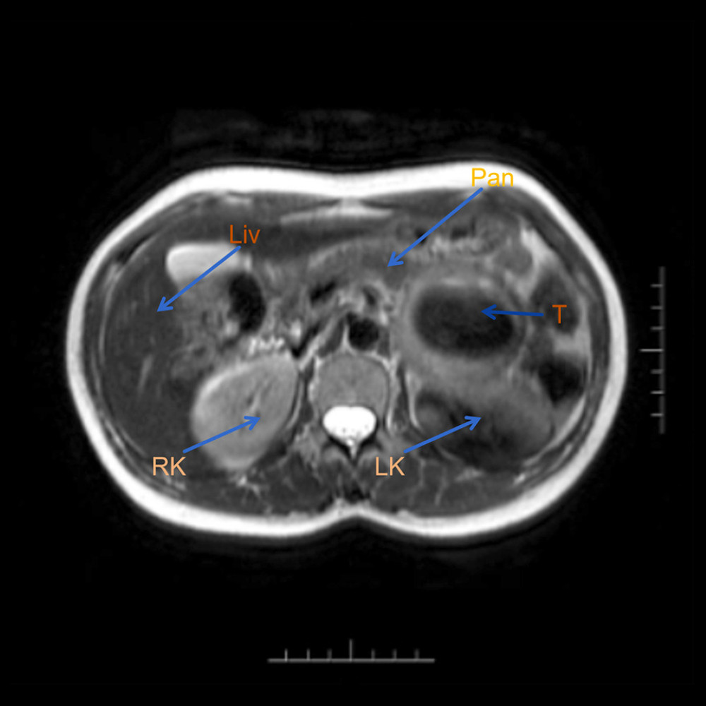
Figure 1
https://jours.isi-science.com/imageXml.php?i=amjcaserep-26-e950574-g001.jpg&idArt=950574&w=1000
Figure 1
Hình 1
Preoperative MRI (RK – right kidney; LK – left kidney; T – tumor; Pan – pancreas; Liv – liver).
Phim chụp cộng hưởng từ (MRI) trước mổ (RK – thận phải; LK – thận trái; T – khối u; Pan – tuyến tụy; Liv – gan).
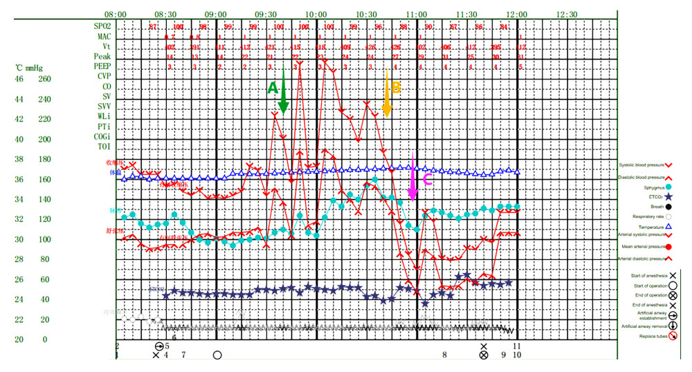
Figure 2
https://jours.isi-science.com/imageXml.php?i=amjcaserep-26-e950574-g002.jpg&idArt=950574&w=1000
Figure 2
Hình 2
Intraoperative anesthesia record. A) Use sodium nitroprusside. B) Use hydrocortisone. C) Use norepinephrine and metaraminol.
Bản ghi gây mê trong mổ. A) Sử dụng natri nitroprusside. B) Sử dụng hydrocortisone. C) Sử dụng norepinephrine và metaraminol.
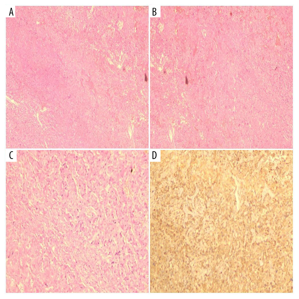
Figure 3
https://jours.isi-science.com/imageXml.php?i=amjcaserep-26-e950574-g003.jpg&idArt=950574&w=1000
Figure 3
Hình 3
Pathology results showing adrenal pheochromocytoma. (A, B) Staining with hematoxylin-eosin (HE); Observation perspective: Two-dimensional planar observation; Internal scale: 1 unit=100–200 μm (low-power). (C) Staining with hematoxylin-eosin (HE); Observation perspective: Two-dimensional planar observation; Internal scale: 1 unit=25–50 μm (high-power). (D) Immunohistochemical staining; Observation perspective: Two-dimensional planar observation; Internal scale: 1 unit=25–50 μm (high-power); Immunohistochemical Results: S100(+), PMS-2 (machine-stained) (+), MLH1 (machine-stained) (focal +), MSH2 (machine-stained) (focal +), MSH6 (machine-stained) (+), β-catenin (membranous +), inhibin-α(−), Ki-67(1%), ATRX(+), SF-1 (+), P27(+), CD56(+), CgA(+), Syn(+), CKL(−), SDHB(+).
Kết quả giải phẫu bệnh cho thấy u tủy thượng thận. (A, B) Nhuộm hematoxylin-eosin (HE); Góc độ quan sát: Quan sát mặt phẳng hai chiều; Thước đo bên trong: 1 đơn vị = 100–200 μm (vật kính nhỏ). (C) Nhuộm hematoxylin-eosin (HE); Góc độ quan sát: Quan sát mặt phẳng hai chiều; Thước đo bên trong: 1 đơn vị = 25–50 μm (vật kính lớn). (D) Nhuộm hóa mô miễn dịch; Góc độ quan sát: Quan sát mặt phẳng hai chiều; Thước đo bên trong: 1 đơn vị = 25–50 μm (vật kính lớn); Kết quả hóa mô miễn dịch: S100(+), PMS-2 (nhuộm máy) (+), MLH1 (nhuộm máy) (+ khu trú), MSH2 (nhuộm máy) (+ khu trú), MSH6 (nhuộm máy) (+), β-catenin (+ ở màng tế bào), inhibin-α(−), Ki-67(1%), ATRX(+), SF-1 (+), P27(+), CD56(+), CgA(+), Syn(+), CKL(−), SDHB(+).
Có thể bạn quan tâm
-
Sarcoidosis thần kinh và sarcoidosis tim đồng mắc: Một lộ trình chẩn đoán đặc biệt
Concomitant Neurosarcoidosis and Cardiac Sarcoidosis: A Peculiar Path to Diagnosis
-
Thay thế bác sĩ bằng điều dưỡng trong môi trường bệnh viện: Hiệu quả đối với kết cục của bệnh nhân, quy trình chăm sóc và chi phí
Nurse-physician substitution in hospital settings: Effects on patient outcomes, care processes, and costs
-
Phối hợp ba thuốc kháng huyết áp liều thấp trong một viên nén sau xuất huyết não
Three Low-Dose Antihypertensive Agents in a Single Pill after Intracerebral Hemorrhage
-
Hantavirus, COVID, norovirus, legionnaires: tại sao tàu du lịch lại dễ bùng phát dịch bệnh đến vậy?
Hantavirus, COVID, norovirus, legionnaires’: why are cruise ships so prone to disease outbreaks?