Ca lâm sàng
Đóng động mạch qua da đối với trường hợp vô tình đặt ống thông vào động mạch dưới đòn trong quá trình đặt catheter tĩnh mạch trung tâm: Báo cáo một ca lâm sàng
Percutaneous Arterial Closure for Inadvertent Subclavian Artery Cannulation During Central Venous Catheterization: A Case Report
Abstract
Tóm tắt
BACKGROUND: Arterial injury during central venous catheterization (CVC) occurs in 0.1% to 2.7% of cases and can lead to serious complications, such as arterial occlusion, embolism, pseudoaneurysm, vessel rupture, and life-threatening hemorrhage. These risks are particularly concerning in critically ill patients, especially those receiving anticoagulation or with acute coagulopathy.
BỐI CẢNH: Tổn thương động mạch trong quá trình đặt catheter tĩnh mạch trung tâm (CVC) xảy ra trong khoảng 0,1% đến 2,7% các trường hợp và có thể dẫn đến các biến chứng nghiêm trọng, chẳng hạn như tắc động mạch, thuyên tắc mạch, giả phình mạch, vỡ mạch máu và xuất huyết đe dọa tính mạng. Những nguy cơ này đặc biệt đáng lo ngại ở những bệnh nhân nguy kịch, đặc biệt là những người đang điều trị bằng thuốc chống đông hoặc bị bệnh lý đông máu cấp tính.
CASE REPORT: We report the case of an 81-year-old woman with a complex medical history, including chronic heart failure, atrial fibrillation, chronic obstructive pulmonary disease (COPD), and chronic kidney disease, whose left subclavian artery was accidentally punctured during central venous catheterization (CVC). The injury was managed successfully by removing the catheter and performing percutaneous arterial closure. The repair involved image-guided percutaneous vascular suturing, using fluoroscopy and real-time contrast imaging. Before the procedure, color Doppler ultrasonography was used to assess the injury’s size and location, while CT angiography evaluated the surrounding vasculature for complications, including arterial rupture or significant bleeding. The postoperative recovery was uneventful, with no signs of pseudoaneurysm or arteriovenous fistula. The patient maintained stable hemodynamics throughout the recovery period and exhibited no signs of further vascular complications.
BÁO CÁO CA LÂM SÀNG: Chúng tôi báo cáo ca lâm sàng của một cụ bà 81 tuổi với tiền sử bệnh lý phức tạp, bao gồm suy tim mạn tính, rung nhĩ, bệnh phổi tắc nghẽn mạn tính (COPD) và bệnh thận mạn tính, người bị đâm nhầm vào động mạch dưới đòn trái trong khi đặt catheter tĩnh mạch trung tâm (CVC). Tổn thương đã được xử trí thành công bằng cách rút catheter và thực hiện đóng động mạch qua da. Quá trình sửa chữa bao gồm khâu mạch máu qua da dưới hướng dẫn hình ảnh, sử dụng màn tăng sáng huỳnh quang và chụp cản quang thời gian thực. Trước khi thực hiện thủ thuật, siêu âm Doppler màu đã được sử dụng để đánh giá kích thước và vị trí tổn thương, trong khi chụp cắt lớp vi tính mạch máu (CTA) được dùng để đánh giá hệ mạch máu xung quanh nhằm tìm các biến chứng, bao gồm vỡ động mạch hoặc chảy máu nghiêm trọng. Quá trình hồi phục sau phẫu thuật diễn ra thuận lợi, không có dấu hiệu của giả phình mạch hoặc rò động tĩnh mạch. Bệnh nhân duy trì huyết động ổn định trong suốt giai đoạn hồi phục và không có dấu hiệu của các biến chứng mạch máu nào khác.
Percutaneous suturing is a safe and effective method for managing mild arterial injuries during central venous catheterization (CVC), especially in patients with complex comorbidities. This approach provides precise hemostasis, minimizes vascular trauma, and offers a minimally invasive solution to prevent significant complications, ensuring a smooth recovery.
Khâu qua da là một phương pháp an toàn và hiệu quả để xử trí các tổn thương động mạch nhẹ trong quá trình đặt catheter tĩnh mạch trung tâm (CVC), đặc biệt ở những bệnh nhân có các bệnh đồng mắc phức tạp. Tiếp cận này giúp cầm máu chính xác, giảm thiểu chấn thương mạch máu và mang lại một giải pháp xâm lấn tối thiểu để ngăn ngừa các biến chứng nghiêm trọng, đảm bảo quá trình hồi phục suôn sẻ.
Keywords: catheterization, central venous, subclavian artery, vascular closure devices
Từ khóa: catheterization, central venous, subclavian artery, vascular closure devices
Introduction
Đặt vấn đề
Central venous catheterization is a routine procedure in clinical practice, commonly used in critical care and therapeutic interventions. Despite its routine nature, complications remain significant and warrant caution 1. Arterial injury, particularly with large-bore catheters, is one of the most frequent complications of this procedure. Delayed recognition of arterial injury can lead to serious outcomes, including pseudoaneurysm, thrombosis, and arteriovenous fistula. Although ultrasound guidance reduces the risk of arterial puncture, arterial injuries may still occur despite its use 2. Subclavian artery (SCA) injury poses unique challenges due to its deep location and non-compressibility, often resulting in ineffective manual hemostasis and increased risk of uncontrolled bleeding 3. In this case, despite the patient’s hypotension and chronic hypoxia, identifying the bleeding source was initially difficult. The deep location of the SCA and the rapid onset of bleeding likely delayed recognition of the injury, as the blood was initially indistinguishable from venous or other hemorrhage sources. Recent advancements in endovascular repair techniques, such as percutaneous vascular suturing devices and covered stents, have emerged as safer and more effective treatment options for complications from arterial punctures 4. This report details an accidental left subclavian artery puncture during central venous catheterization, which was successfully managed through catheter removal followed by percutaneous arterial suturing, preventing further complications. This case highlights the potential of endovascular therapy in managing arterial punctures, particularly when conventional methods fail to achieve adequate hemostasis, providing a minimally invasive and safe alternative. By examining the broader application of percutaneous vascular suturing devices in similar cases, this case aims to provide practical guidance for managing such events and promote the clinical adoption of endovascular techniques.
Đặt catheter tĩnh mạch trung tâm là một thủ thuật thường quy trong thực hành lâm sàng, thường được sử dụng trong hồi sức tích cực và các can thiệp điều trị. Dù là một thủ thuật thường quy, các biến chứng vẫn ở mức đáng kể và đòi hỏi sự thận trọng 1. Tổn thương động mạch, đặc biệt là với các ống thông lòng lớn, là một trong những biến chứng thường gặp nhất của thủ thuật này. Việc nhận biết muộn tổn thương động mạch có thể dẫn đến những hậu quả nghiêm trọng, bao gồm giả phình mạch, huyết khối và rò động tĩnh mạch. Mặc dù việc hướng dẫn bằng siêu âm làm giảm nguy cơ đâm nhầm vào động mạch, tổn thương động mạch vẫn có thể xảy ra bất chấp việc sử dụng phương pháp này 2. Tổn thương động mạch dưới đòn (SCA) đặt ra những thách thức độc sảo do vị trí nằm sâu và tính chất không thể ép lòng mạch, thường dẫn đến việc ép cầm máu bằng tay không hiệu quả và làm tăng nguy cơ chảy máu không kiểm soát 3. Trong trường hợp này, bất chấp tình trạng tụt huyết áp và thiếu oxy mạn tính của bệnh nhân, việc xác định nguồn chảy máu ban đầu gặp nhiều khó khăn. Vị trí nằm sâu của động mạch dưới đòn và sự khởi phát chảy máu nhanh chóng có thể đã làm chậm trễ việc nhận biết tổn thương, do máu ban đầu không thể phân biệt được với máu tĩnh mạch hoặc các nguồn xuất huyết khác. Những tiến bộ gần đây trong kỹ thuật sửa chữa nội mạch, chẳng hạn như các thiết bị khâu mạch máu qua da và giá đỡ mạch máu có màng bọc (stent graft), đã nổi lên như những lựa chọn điều trị an toàn và hiệu quả hơn cho các biến chứng từ việc đâm nhầm động mạch 4. Báo cáo này chi tiết về một trường hợp đâm nhầm vào động mạch dưới đòn trái trong khi đặt catheter tĩnh mạch trung tâm, tổn thương này đã được xử trí thành công bằng cách rút catheter kết hợp với khâu động mạch qua da, ngăn ngừa các biến chứng nặng hơn. Ca lâm sàng này làm nổi bật tiềm năng của liệu pháp nội mạch trong việc xử trí các trường hợp đâm nhầm động mạch, đặc biệt khi các phương pháp thông thường thất bại trong việc đạt được cầm máu thích hợp, mang lại một giải pháp thay thế xâm lấn tối thiểu và an toàn. Bằng cách xem xét việc áp dụng rộng rãi hơn các thiết bị khâu mạch máu qua da trong các trường hợp tương tự, ca lâm sàng này nhằm mục đích cung cấp hướng dẫn thực hành cho việc xử trí các biến cố như vậy và thúc đẩy việc ứng dụng kỹ thuật nội mạch trong lâm sàng.
Case Report
Báo cáo ca lâm sàng
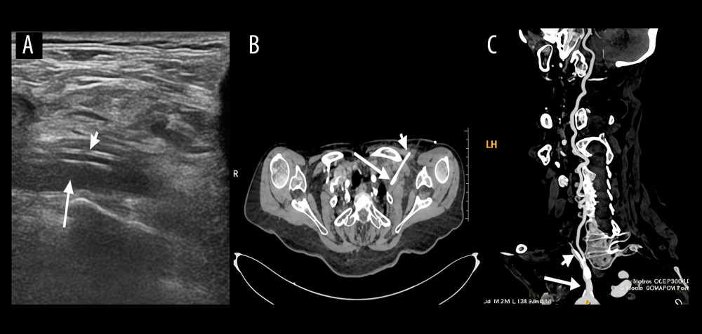
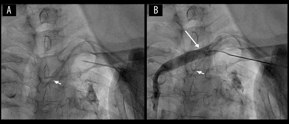
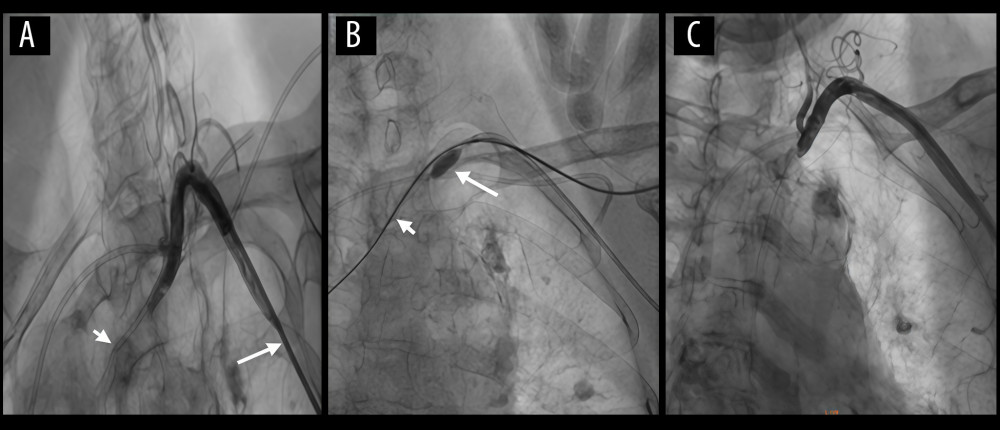
The patient was an 81-year-old woman with a 20-year history of recurrent palpitations. Recently, her palpitations had worsened, accompanied by dizziness and nausea. She had multiple comorbidities, including chronic heart failure, paroxysmal atrial fibrillation, hypertension, type 2 diabetes mellitus, chronic obstructive pulmonary disease (COPD), and chronic kidney disease (CKD). Over 20 years ago, she underwent radiofrequency ablation for atrial fibrillation and has been on long-term Edoxaban Tosylate therapy. A second ablation was performed five years ago for recurrent atrial fibrillation, which led to symptomatic improvement. Physical examination revealed hypotension (68/57 mmHg), tachycardia (120 bpm), and mild bilateral lower limb edema. Laboratory tests showed anemia (hemoglobin 99 g/L), elevated serum creatinine (230 μmol/L), and proteinuria (1+). Electrocardiography revealed 2: 1 atrial flutter, and echocardiography demonstrated left ventricular hypertrophy, biatrial enlargement, pulmonary hypertension (PASP ~86 mmHg), and moderate tricuspid regurgitation. Due to hypotension and inadequate peripheral venous access, a left subclavian vein catheterization was performed to improve fluid administration. The skin was disinfected, a sterile drape was applied, and local anesthesia was administered. The puncture needle was inserted at a 30°angle toward the sternal notch. Dark red blood was aspirated and mistakenly identified as venous. A guidewire and deep venous catheter were subsequently inserted. After the procedure, a mild pulsatile blood flow was observed slowly refluxing within the catheter. Arterial blood gas analysis showed a PaO2 of 68 mmHg, raising suspicion of catheter misplacement into the left subclavian artery. Emergent cervical vascular ultrasonography and contrast-enhanced CT confirmed that the catheter was in the left subclavian artery, with its tip at the arterial origin. No arteriovenous fistula was observed (Figure 1A–1C). Given the deep anatomical location of the left subclavian artery and its non-compressibility, along with the patient’s overall condition, percutaneous arterial suturing was selected for vascular repair. A new left subclavian vein catheterization was conducted before the procedure. Left subclavian vein angiography was performed, with no contrast agent leakage observed (Figure 2A, 2B). After routine disinfection, sterile draping, and local anesthesia, successful puncture of the left radial artery was achieved. A 7F radial artery sheath was placed, and under fluoroscopic guidance, a JR4.0 guiding catheter was advanced to the proximal segment of the left subclavian artery. Angiography revealed the central venous catheter traversing the left subclavian artery without significant contrast extravasation. A guidewire was advanced through the injured arterial segment into the ascending aorta. A 5.0×8 mm NC TREK balloon was placed proximal to the injury site over the guidewire and continuously inflated to occlude blood flow. A 6F ExoSeal (Cordis) vascular closure device was deployed through the misplaced catheter, sealing the puncture site. Gradual withdrawal of the device was performed while monitoring bleeding from the indicator tube. When bleeding ceased, indicating that the device tip had reached the correct extravascular position adjacent to the arterial wall, the occlusion mechanism was deployed. The device was then removed, and gentle manual pressure was applied to the puncture site. Follow-up angiography demonstrated intact flow in the left subclavian artery, with no contrast extravasation (Figure 3A–3C). The procedure was completed, and the patient was transferred to the cardiac intensive care unit (CICU). Postoperatively, the patient recovered well without complications such as pseudoaneurysm, arterial dissection, or vascular occlusion.
Bệnh nhân là một cụ bà 81 tuổi với tiền sử 20 năm bị hồi hộp đánh trống ngực tái phát. Gần đây, tình trạng hồi hộp đánh trống ngực của bà trở nên trầm trọng hơn, kèm theo chóng mặt và buồn nôn. Bà có nhiều bệnh đồng mắc, bao gồm suy tim mạn tính, rung nhĩ kịch phát, tăng huyết áp, đái tháo đường típ 2, bệnh phổi tắc nghẽn mạn tính (COPD) và bệnh thận mạn tính (CKD). Hơn 20 năm trước, bà đã được triệt đốt rung nhĩ bằng năng lượng sóng có tần số radio và đã điều trị dài hạn bằng Edoxaban Tosylate. Cách đây 5 năm, một lần triệt đốt thứ hai đã được thực hiện do rung nhĩ tái phát, giúp cải thiện triệu chứng. Thăm khám lâm sàng ghi nhận tình trạng tụt huyết áp (68/57 mmHg), nhịp tim nhanh (120 chu kỳ/phút) và phù nhẹ hai chi dưới. Các xét nghiệm cận lâm sàng cho thấy tình trạng thiếu máu (hemoglobin 99 g/L), nồng độ creatinine huyết thanh tăng cao (230 μmol/L) và protein niệu (1+). Điện tâm đồ ghi nhận cuồng nhĩ dẫn truyền 2:1, và siêu âm tim cho thấy phì đại thất trái, giãn hai tâm nhĩ, tăng áp lực động mạch phổi (PASP ~86 mmHg) và hở van ba lá mức độ trung bình. Do tình trạng tụt huyết áp và đường truyền tĩnh mạch ngoại vi không đảm bảo, thủ thuật đặt catheter tĩnh mạch dưới đòn trái đã được thực hiện để tăng cường truyền dịch. Da vùng bẹn/cổ được sát khuẩn, trải săng vô khuẩn và tiến hành gây tê tại chỗ. Kim chọc dò được đâm một góc 30° hướng về phía khuyết ức. Máu đỏ sẫm được hút ra và bị nhận định nhầm là máu tĩnh mạch. Một dây dẫn (guidewire) và một catheter tĩnh mạch sâu sau đó đã được luồn vào. Sau thủ thuật, dòng máu đập nhẹ theo nhịp đập được quan sát thấy đang trào ngược chậm trong lòng catheter. Kết quả khí máu động mạch cho thấy PaO2 là 68 mmHg, làm dấy lên nghi ngờ về việc đặt nhầm vị trí catheter vào động mạch dưới đòn trái. Siêu âm mạch máu cổ cấp cứu và chụp cắt lớp vi tính (CT) có cản quang xác nhận catheter nằm trong động mạch dưới đòn trái, với đầu catheter nằm ở gốc động mạch. Không quan sát thấy rò động tĩnh mạch (Figure 1A–1C). Do vị trí giải phẫu nằm sâu của động mạch dưới đòn trái và tính chất không thể ép lòng mạch, cùng với tình trạng tổng thể của bệnh nhân, phương pháp khâu động mạch qua da đã được lựa chọn để sửa chữa mạch máu. Một thủ thuật đặt catheter tĩnh mạch dưới đòn trái mới đã được tiến hành trước khi thực hiện ca can thiệp. Chụp mạch máu tĩnh mạch dưới đòn trái được thực hiện, không ghi nhận tình trạng rò rỉ thuốc cản quang (Figure 2A, 2B). Sau khi sát khuẩn thường quy, trải săng vô khuẩn và gây tê tại chỗ, việc chọc dò động mạch quay trái đã thành công. Một ống mở đường vào (sheath) động mạch quay kích cỡ 7F được đặt vào, và dưới hướng dẫn của màn tăng sáng huỳnh quang, một catheter dẫn đường (guiding catheter) JR4.0 được đưa đến đoạn gần của động mạch dưới đòn trái. Kết quả chụp mạch cho thấy catheter tĩnh mạch trung tâm đi xuyên qua động mạch dưới đòn trái mà không có sự thoát mạch thuốc cản quang đáng kể. Một dây dẫn được đưa qua đoạn động mạch bị tổn thương vào động mạch chủ lên. Một bóng NC TREK kích thước 5.0×8 mm được đặt ở đoạn gần vị trí tổn thương qua dây dẫn và được bơm phồng liên tục để chặn dòng máu. Một thiết bị đóng mạch máu ExoSeal (Cordis) kích cỡ 6F được triển khai qua ống catheter bị đặt nhầm vị trí, bít kín vị trí chọc dò. Quá trình rút dần thiết bị được thực hiện trong khi theo dõi tình trạng chảy máu từ ống chỉ thị. Khi máu ngừng chảy, cho thấy đầu thiết bị đã đạt đến vị trí chính xác ngoài mạch máu sát với thành động mạch, cơ chế bít kín được kích hoạt. Thiết bị sau đó được rút ra, và lực ép nhẹ bằng tay được áp dụng lên vị trí chọc dò. Chụp mạch kiểm tra sau đó cho thấy dòng chảy trong động mạch dưới đòn trái thông suốt, không có hiện tượng thoát mạch thuốc cản quang (Figure 3A–3C). Thủ thuật hoàn thành, và bệnh nhân được chuyển đến đơn vị hồi sức tim mạch sâu (CICU). Sau can thiệp, bệnh nhân phục hồi tốt và không gặp phải các biến chứng như giả phình mạch, bóc tách động mạch hay tắc mạch.
Discussion
Bàn luận
Despite significant progress in imaging-guided techniques, arterial injury remains a well-recognized complication of central venous catheterization. Although relatively uncommon, such events can result in severe and potentially life-threatening consequences. Subclavian artery (SCA) injuries are particularly challenging owing to the vessel’s deep anatomical course and lack of compressibility, which limit the effectiveness of conventional hemostatic maneuvers. These anatomical and technical factors underscore the need for prompt recognition and effective management to minimize morbidity and mortality 5.
Mặc dù đã có những tiến bộ đáng kể trong các kỹ thuật dưới hướng dẫn hình ảnh, tổn thương động mạch vẫn là một biến chứng đã được ghi nhận rõ ràng của thủ thuật đặt catheter tĩnh mạch trung tâm. Dù tương đối ít gặp, những biến cố như vậy có thể dẫn đến các hậu quả nghiêm trọng và tiềm ẩn nguy cơ đe dọa tính mạng. Tổn thương động mạch dưới đòn (SCA) đặc biệt mang tính thách thức do đường đi giải phẫu nằm sâu của mạch máu và tính chất không thể ép lòng mạch, điều này làm hạn chế hiệu quả của các biện pháp ép cầm máu thông thường. Những yếu tố giải phẫu và kỹ thuật này nhấn mạnh nhu cầu cần phải nhận biết nhanh chóng và xử trí hiệu quả nhằm giảm thiểu tỷ lệ mắc bệnh và tử vong 5.
This case highlights the complexity of managing an SCA injury in a high-risk patient with multiple comorbidities, including chronic heart failure, atrial fibrillation, and chronic kidney disease. The combination of deep vascular anatomy and hemodynamic instability increased the likelihood of inadvertent arterial puncture and delayed recognition. Nevertheless, rapid diagnosis and timely intervention facilitated successful management, preventing secondary complications such as hematoma formation or pseudoaneurysm development 6.
Ca lâm sàng này làm nổi bật tính phức tạp của việc xử trí tổn thương động mạch dưới đòn ở một bệnh nhân có nguy cơ cao với nhiều bệnh đồng mắc, bao gồm suy tim mạn tính, rung nhĩ và bệnh thận mạn tính. Sự kết hợp giữa cấu trúc giải phẫu mạch máu nằm sâu và tình trạng huyết động không ổn định đã làm tăng khả năng vô tình đâm nhầm vào động mạch và làm chậm trễ việc nhận biết. Mặc dù vậy, việc chẩn đoán nhanh chóng và can thiệp kịp thời đã tạo điều kiện cho việc xử trí thành công, ngăn ngừa các biến chứng thứ phát như hình thành khối tụ máu hoặc phát triển giả phình mạch 6.
Traditional management of arterial injuries from CVC includes manual compression, endovascular interventions, and open surgical repair. While percutaneous stent graft implantation is commonly used in such cases, newer approaches, such as percutaneous arterial suturing, provide an effective and minimally invasive alternative, particularly for minor arterial injuries in anatomically challenging regions, like the SCA 7. Endovascular repair has become increasingly favored due to its minimally invasive nature; however, percutaneous suturing allows for precise closure of the arterial defect with minimal tissue trauma and a faster recovery 8.
Xử trí truyền thống đối với các tổn thương động mạch do CVC bao gồm ép bằng tay, can thiệp nội mạch và phẫu thuật sửa chữa mở. Trong khi phương pháp đặt giá đỡ mạch máu có màng bọc (stent graft) qua da thường được sử dụng trong các trường hợp này, các tiếp cận mới hơn, chẳng hạn như khâu động mạch qua da, mang lại một giải pháp thay thế hiệu quả và xâm lấn tối thiểu, đặc biệt đối với các tổn thương động mạch nhỏ ở những vùng có cấu trúc giải phẫu đầy thách thức như động mạch dưới đòn 7. Sửa chữa nội mạch ngày càng được ưa chuộng do tính chất xâm lấn tối thiểu của nó; tuy nhiên, khâu qua da cho phép đóng chính xác tổn thương khuyết tật động mạch với chấn thương mô tối thiểu và thời gian phục hồi nhanh hơn 8.
In the present case, percutaneous suturing was selected, given the limited extent of arterial injury and the patient’s frail condition. The method provided accurate closure with minimal vascular manipulation, offering an optimal balance between efficacy and safety 9. Imaging played an indispensable role throughout diagnosis and treatment. Preoperative color Doppler ultrasonography and CT angiography accurately localized the puncture site and delineated the extent of vascular injury, while intraoperative digital subtraction angiography and fluoroscopic guidance provided real-time visualization that enhanced procedural precision and ensured complete hemostasis 10.
Trong trường hợp này, phương pháp khâu qua da đã được lựa chọn do mức độ tổn thương động mạch hạn chế và tình trạng thể trạng suy kiệt của bệnh nhân. Phương pháp này giúp đóng chính xác với sự thao tác mạch máu tối thiểu, mang lại sự cân bằng tối ưu giữa tính hiệu quả và tính an toàn 9. Chẩn đoán hình ảnh đóng một vai trò không thể thiếu trong suốt quá trình chẩn đoán và điều trị. Siêu âm Doppler màu và chụp cắt lớp vi tính mạch máu (CTA) trước phẫu thuật đã xác định chính xác vị trí chọc dò và mô tả mức độ tổn thương mạch máu, trong khi chụp mạch số hóa xóa nền (DSA) và hướng dẫn bằng màn tăng sáng huỳnh quang trong phẫu thuật đã cung cấp hình ảnh trực quan theo thời gian thực giúp nâng cao độ chính xác của thủ thuật và đảm bảo cầm máu hoàn toàn 10.
This case highlights the importance of accurate diagnosis and timely intervention in managing inadvertent arterial punctures during CVC. The use of percutaneous arterial suturing, combined with advanced imaging techniques, offers a safe and effective approach, particularly in high-risk patients with complex comorbidities. As technology and devices continue to evolve, these minimally invasive methods are expected to improve, allowing for even greater precision in managing such complications.
Ca lâm sàng này nhấn mạnh tầm quan trọng của việc chẩn đoán chính xác và can thiệp kịp thời trong việc xử trí các trường hợp vô tình đâm nhầm động mạch trong quá trình đặt CVC. Việc sử dụng phương pháp khâu động mạch qua da, kết hợp với các kỹ thuật hình ảnh tiên tiến, mang lại một tiếp cận an toàn và hiệu quả, đặc biệt ở những bệnh nhân nguy cơ cao có các bệnh đồng mắc phức tạp. Khi công nghệ và các thiết bị tiếp tục phát triển, các phương pháp xâm lấn tối thiểu này được kỳ vọng sẽ cải thiện hơn nữa, cho phép đạt độ chính xác cao hơn nữa trong việc xử trí các biến chứng như vậy.
Conclusions
Kết luận
This case illustrates the successful management of an inadvertent perforation of the left subclavian artery during central venous catheterization. Percutaneous arterial repair using a vascular closure device avoided the need for open surgery and minimized postoperative complications. Minimally invasive techniques, especially in cases of deep arterial injuries where manual compression is ineffective, have shown favorable outcomes. Key clinical lessons include the importance of timely recognition and the role of percutaneous techniques in managing complex arterial injuries.
Ca lâm sàng này minh họa cho việc xử trí thành công một trường hợp vô tình làm thủng động mạch dưới đòn trái trong quá trình đặt catheter tĩnh mạch trung tâm. Việc sửa chữa động mạch qua da bằng thiết bị đóng mạch máu đã tránh được việc phải phẫu thuật mở và giảm thiểu các biến chứng sau phẫu thuật. Các kỹ thuật xâm lấn tối thiểu, đặc biệt là trong các trường hợp tổn thương động mạch sâu nơi mà phương pháp ép bằng tay không hiệu quả, đã cho thấy những kết quả khả quan. Các bài học lâm sàng chính bao gồm tầm quan trọng của việc nhận biết kịp thời và vai trò của các kỹ thuật qua da trong việc xử trí các tổn thương động mạch phức tạp.
Reference
- Kusminsky RE, Complications of central venous catheterization: J Am Coll Surg, 2007; 204(4); 681-96
- McGee DC, Gould MK, Preventing complications of central venous catheterization: N Engl J Med, 2003; 348(12); 1123-33
- Guilbert MC, Elkouri S, Bracco D, Arterial trauma during central venous catheter insertion: Case series, review and proposed algorithm: J Vasc Surg, 2008; 48(5); 918-25
- Thalhammer C, Kirchherr AS, Uhlich F, Postcatheterization pseudoaneurysms and arteriovenous fistulas: Repair with percutaneous implantation of endovascular covered stents: Radiology, 2000; 214(1); 127-31
- Dixon OGB, Smith GE, Carradice D, Chetter IC, A systematic review of management of inadvertent arterial injury during central venous catheterisation: J Vasc Access, 2017; 18(6); 495-502
- Branco BC, Boutrous ML, DuBose JJ, Outcomes after subclavian and axillary artery injuries: A National Trauma Data Bank analysis: J Vasc Surg, 2016; 63(3); 702-9
- Pikwer A, Acosta S, Kölbel T, Management of inadvertent arterial catheterisation associated with central venous access procedures: Eur J Vasc Endovasc Surg, 2009; 38; 707-14
- Trabattoni D, Bartorelli AL, Agrifoglio M, Montorsi P, Endovascular repair of iatrogenic subclavian artery perforations using the Hemobahn stent-graft: J Endovasc Ther, 2001; 8; 417-21
- AlSheikh S, Aldossary MY, Najeeb R, Management of arterial trauma during central venous catheter insertion using a percutaneous suture-mediated closure device (Perclose ProGlide): A report of two cases and literature review: Signa Vitae, 2023; 19; 211-16
- Fritz J, Efron DT, Fishman EK, Multidetector CT and three-dimensional CT angiography of upper extremity arterial injury: Emerg Radiol, 2015; 22; 45-52
Có thể bạn quan tâm
-
Viêm mạch u hạt tăng bạch cầu ái toan âm tính với ANCA biểu hiện bằng hẹp tá tràng: Một báo cáo ca lâm sàng
ANCA-Negative Eosinophilic Granulomatosis With Polyangiitis Presenting With Duodenal Stenosis: A Case Report
-
Lợi ích và tác hại của các loại thuốc từ cần sa đối với chứng đau thần kinh mạn tính ở người lớn
Benefits and harms of cannabis-based medicines for chronic neuropathic pain in adults
-
Khi định nghĩa về tự kỷ mở rộng, liệu chúng ta có đang bỏ quên những người có nhu cầu lớn nhất không?
As the definition of autism expands, are we losing sight of those with the greatest needs?