Miễn phí
Tụ mủ dưới màng cứng và tràn khí nội sọ do viêm xoang polyp mạn tính ở một phụ nữ lớn tuổi có sự phục hồi thần kinh nhanh chóng: Báo cáo một ca bệnh hiếm gặp
Subdural Empyema and Pneumocephalus From Chronic Polypoid Sinusitis in an Elderly Woman With Rapid Neurological Recovery: A Report of a Rare Case
John Martinez; Elizabeth Nagidi; Mark Rigby; Lina Ataya; Aftab Macksood; Ghiath Bayasi; Kelsee Rotondo. American Journal of Case Reports. doi: 10.12659/AJCR.949999. Open access.
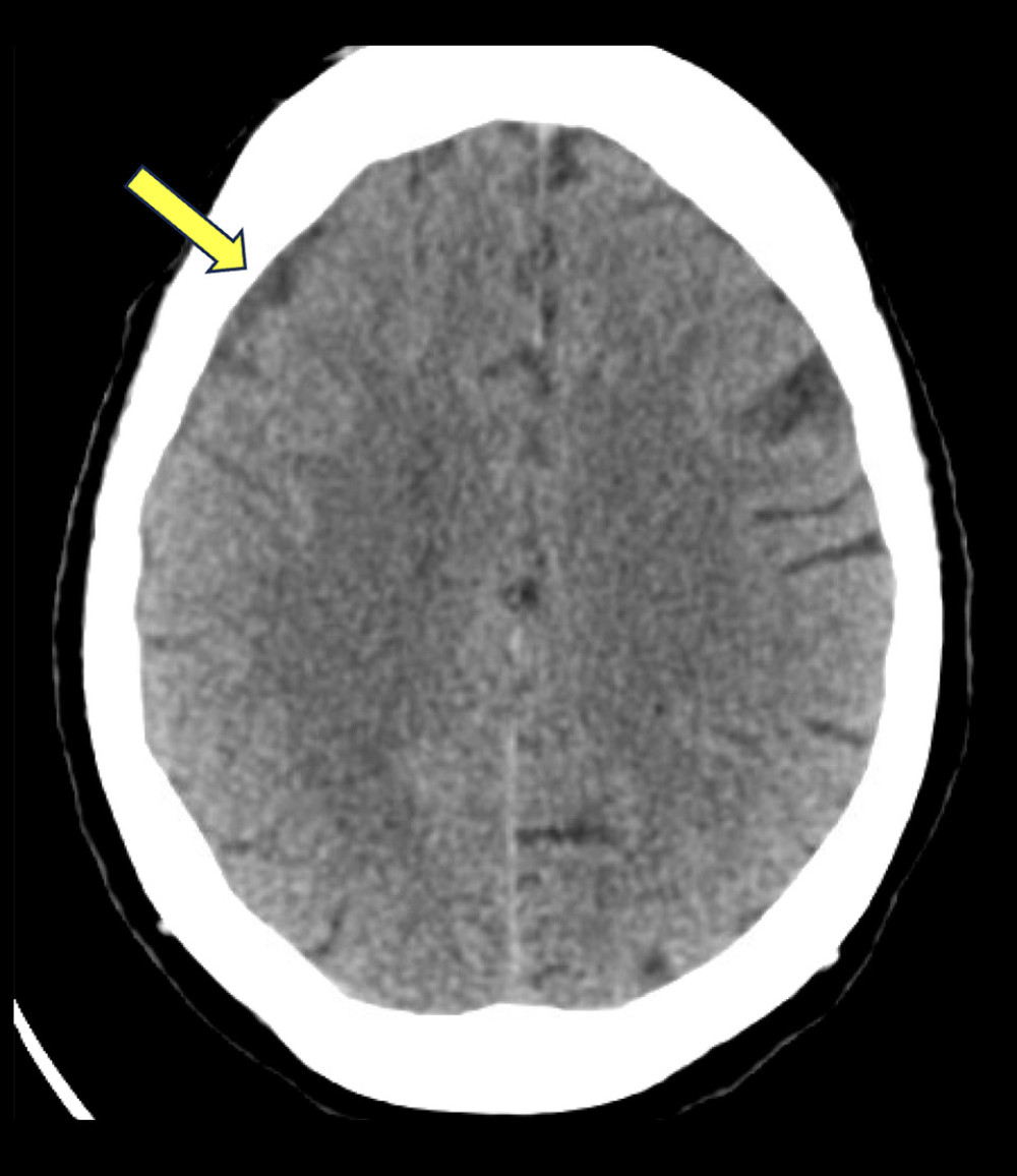
Figure 1
https://jours.isi-science.com/imageXml.php?i=amjcaserep-26-e949999-g001.jpg&idArt=949999&w=1000
Figure 1
Hình 1
Increased density in the extra-axial space is seen on initial CT brain without contrast, most commonly seen in subdural hematoma.
Tăng đậm độ trong khoang ngoài trục được quan sát thấy trên phim chụp CT não không cản quang ban đầu, thường gặp nhất trong tụ máu dưới màng cứng.
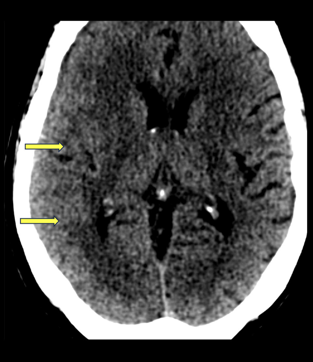
Figure 2
https://jours.isi-science.com/imageXml.php?i=amjcaserep-26-e949999-g002.jpg&idArt=949999&w=1000
Figure 2
Hình 2
Abnormal soft tissue is observed in the brain on the initial CT scan of the brain without contrast. Sulcal effacement is seen in the right posterior temporal and occipital regions.
Mô mềm bất thường được quan sát thấy trong não trên phim chụp CT não không cản quang ban đầu. Hiện tượng xóa các rãnh não được ghi nhận ở vùng thái dương sau và vùng chẩm bên phải.
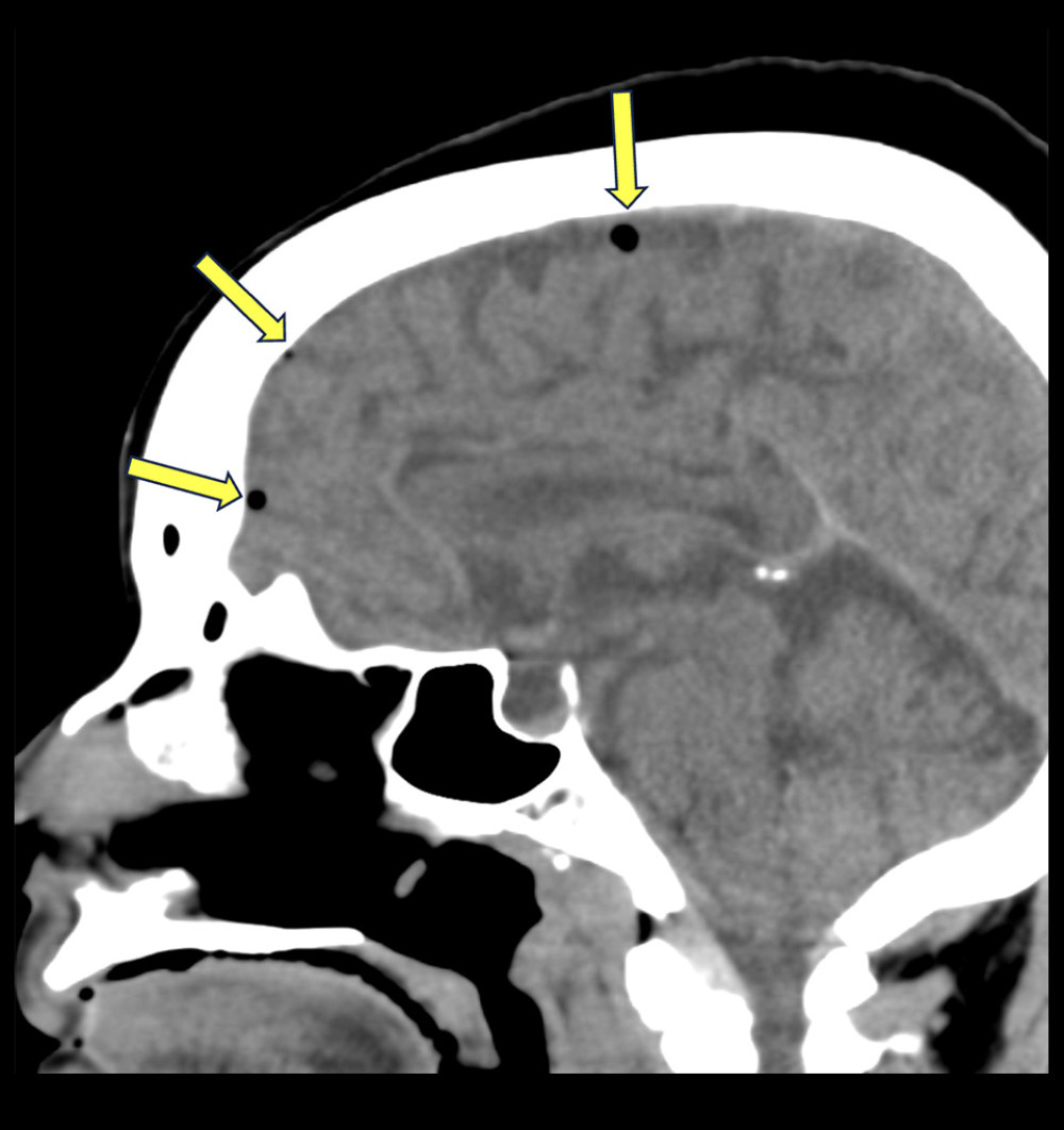
Figure 3
https://jours.isi-science.com/imageXml.php?i=amjcaserep-26-e949999-g003.jpg&idArt=949999&w=1000
Figure 3
Hình 3
Pneumocephalus is visible in the extra-axial space on the initial CT scan of the brain without contrast, most often due to venous air embolus.
Tràn khí nội sọ có thể nhìn thấy trong khoang ngoài trục trên phim chụp CT não không cản quang ban đầu, thường do thuyên tắc khí tĩnh mạch.
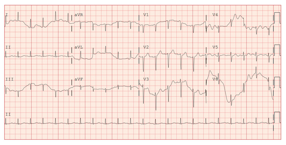
Figure 4
https://jours.isi-science.com/imageXml.php?i=amjcaserep-26-e949999-g004.jpg&idArt=949999&w=1000
Figure 4
Hình 4
EKG showed sinus tachycardia at 128 beats per minute (bpm) without ischemic changes.
Điện tâm đồ (EKG) cho thấy nhịp xoang nhanh ở mức 128 chu kỳ/phút (bpm) và không có thay đổi thiếu máu cục bộ.
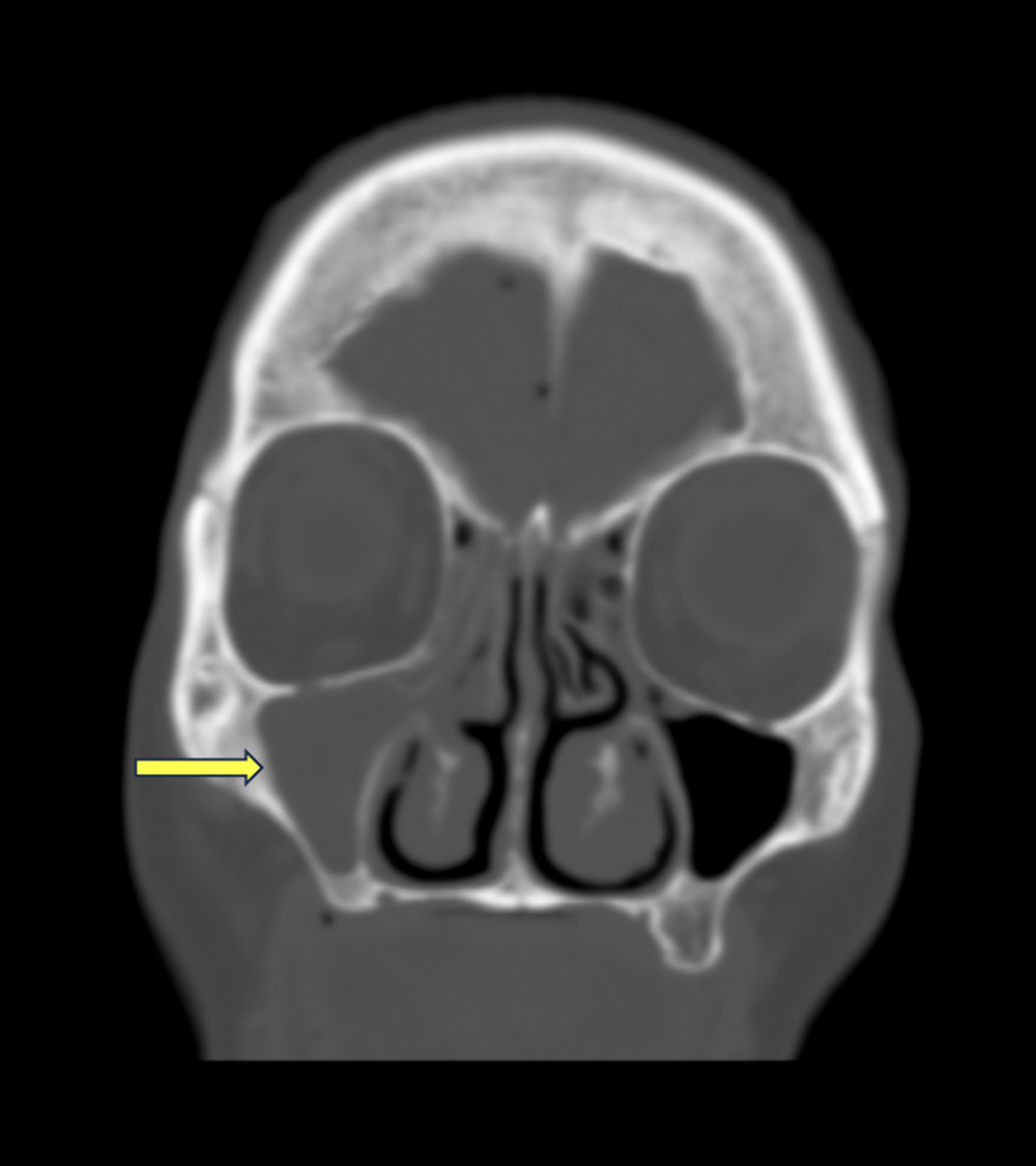
Figure 5
https://jours.isi-science.com/imageXml.php?i=amjcaserep-26-e949999-g005.jpg&idArt=949999&w=1000
Figure 5
Hình 5
Complete opacification of the right maxillary sinus is seen on initial CT of the brain without contrast. Mucosal thickening and right sinus hypoplasia are also seen.
Mờ hoàn toàn xoang hàm bên phải được quan sát thấy trên phim chụp CT não không cản quang ban đầu. Tình trạng dày niêm mạc và kém phát triển xoang bên phải cũng được ghi nhận.
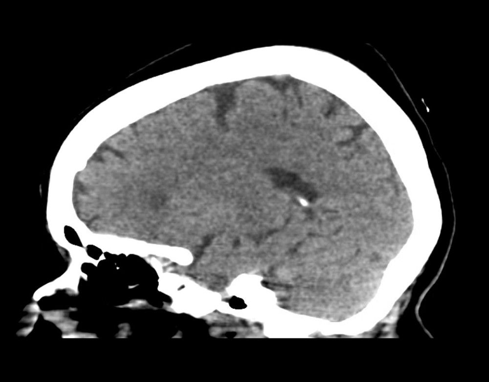
Figure 6
https://jours.isi-science.com/imageXml.php?i=amjcaserep-26-e949999-g006.jpg&idArt=949999&w=1000
Figure 6
Hình 6
CT brain after right frontal craniotomy without contrast. Interval resolution of previously present small-volume right frontal pneumocephalus.
Chụp CT não không cản quang sau phẫu thuật mở hộp sọ trán phải. Ghi nhận sự thoái triển theo thời gian của tình trạng tràn khí vùng trán phải thể tích nhỏ đã xuất hiện trước đó.
Có thể bạn quan tâm
-
Bệnh giảm gammaglobulin máu Bruton: Chuỗi ca bệnh và tổng quan tài liệu từ Bệnh viện Trung ương King Fahad, Jizan, Ả Rập Xê Út
Bruton’s Agammaglobulinemia: Case Series and Literature Review From King Fahad Central Hospital, Jizan, Saudi Arabia
-
Phối hợp ba thuốc kháng huyết áp liều thấp trong một viên nén sau xuất huyết não
Three Low-Dose Antihypertensive Agents in a Single Pill after Intracerebral Hemorrhage
-
Những gì Canada, Vương quốc Anh và các quốc gia G7 khác đã học được về việc xây dựng hệ thống giáo dục kiên cường trong đại dịch COVID-19
What Canada, the U.K. and other G7 nations learned about building resilient education systems during the COVID-19 pandemic